Necrotizing Enterocolitis
Necrotizing Enterocolitis. Jennifer Huesgen N7320 Advanced Pediatric Physiology November 23, 2010. Necrotizing Enterocolitis (NEC). Necrotizing Enterocolitis (NEC) is a serious gastrointestinal disorder that affects preterm and newborn infants.

Necrotizing Enterocolitis
E N D
Presentation Transcript
Necrotizing Enterocolitis Jennifer Huesgen N7320 Advanced Pediatric Physiology November 23, 2010
Necrotizing Enterocolitis (NEC) • Necrotizing Enterocolitis (NEC) is a serious gastrointestinal disorder that affects preterm and newborn infants. • NEC is the leading cause of neonatal morbidity and mortality (Hunter, Upperman, Ford, and Camerini, 2008). • The cause of NEC is not well known, which makes prevention difficult. • Risk factors include premature birth, enteral feedings, and bacterial colonization (Hunter et al., 2008).
NEC • “Necrotizing Enterocolitis is an ischemic, inflammatory condition of the bowel that causes necrosis, perforation, and death if untreated,” (McCance & Huether, 2010, p. 1529). • NEC is the result of a severe inflammatory process that predisposes the tissue to infection and can lead to intestinal necrosis (Carter, 2007). • The exact pathology of NEC is largely unknown.
Risk Factors • There are three risk factors that are believed to predispose an infant to NEC • There must be damage to the intestinal mucosa. • Bacteria must colonize the gastrointestinal tract. • There must be a substrate for fermentation by the bacteria, such as enteral feedings. (Con Yost, 2005). Another common risk factor is premature birth (Hunter et al., 2008).
Damage to the mucosa • Intestinal hypoxia may result from pulmonary immaturity or overwhelming sepsis with hypotension, which can lead to decreased intestinal blood flow (Con Yost, 2005). • The body will conserve the blood flow to vital organs, such as the brain and heart, and shunt flow from non-vital organs, such as the intestines (Gregory, 2008). • The decreased blood flow causes ischemia, which can lead to the development of NEC (Gregory, 2008).
Epithelial Mucosa • The intestinal epithelium acts as a barrier between the body and the intestinal tract (Hunter et al., 2008). • The epithelial cells provide a physical barrier (Hunter et al., 2008). • Goblet cells in the epithelial layer produce mucus made of glycoproteins called mucins (Hunter et al., 2008) • A deficiency in mucus production can allow bacteria to enter the epithelium and start the process for NEC (Hunter et al., 2008).
Epithelial Mucosa • Tight junctions between cells contribute to the semipermeable properties of the cells (Hunter et al., 2008) • Immaturity of the tight junctions can lead to increased permeability of the gut (Hunter et al., 2008). • When bacteria is introduced, it can cause the release of cytokines that disrupt the tight junctions and allow translocation of bacteria and other products (Hunter et al, 2008). • Inflammatory cytokines disrupt the epithelial barrier through overproduction of nitric oxide (NO) and results in cell death (Hunter et al., 2008). • NO causes vasodilatation and increases vascular resistance which decreases blood flow and oxygen, resulting in hypoxia (Gibbs, Lin, & Holzman, 2007).
Epithelial Mucosa • “Immaturity of the intestinal mechanical and biochemical barrier contributes to increased permeability resulting in translocation of bacteria and other substances, causing injury, inflammation, and development of systemic inflammatory disease,” (McCance & Huether, 2010, p. 1529).
Epithelial Mucosa and the Inflammatory Response • The gastrointestinal tract is composed of lymphoid tissue (Hunter et al., 2008). • Macrophages present antigens to lymphocytes. Yet, in newborns, this process is less efficient and the body is less able to detect and respond to pathogens (Hunter et al., 2008). • IgA inhibits bacterial and viral attachment in the mucosa. In infants, there is a deficiency of IgA, which increases the risk of infection (Hunter et al., 2008). • B and T cells are immature and less responsive to stimuli (Hunter et al., 2008).
Presence of Bacteria • Premature infants may have an unfavorable balance between commensal and pathogenic bacteria (McCance & Heuther, 2010). • The newborn intestine has no bacterial flora, but is colonized by the mother at birth, which provides protection from pathogens (Hunter et al., 2008). • No single bacteria or virus has been isolated in NEC, but Enterobacteriaceae, Clostridia, and Staphylococcus were commonly identified (Hunter et al., 2008). • Infants also lack anaerobic species, such as Lactobacillus, which creates an environment for overgrowth of bacteria and initiation of inflammatory cascade (Con Yost, 2005).
Enteral Feedings • “The overgrowth of pathogens in both the small and large intestine is thought to be promoted by unabsorbed luminal nutrient, which acts as a substrate for bacterial growth,” (Gregory, 2008,p. 262). • The bacteria ferments, which produces intraluminal gas. The gas results in distention and increased pressure, which decreases blood flow (Gregory, 2008). • NEC occurs more frequently in infants fed formula than those fed breastmilk (Con Yost, 2005). • Breast milk increases the diversity of the GI tract and contains immunomodulatory factors (Con Yost, 2005).
No embryologic or genetic influences Premature infants are more prone to NEC due to their underdeveloped mucosa and immune response, as previously described. There is a possibility that the hypoxic insult could occur in utero, but that would not be related to embryologic development of the fetus.
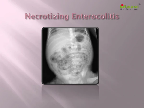
Clinical Manifestations • Symptoms usually present two weeks after birth, with an earlier presentation in full-term infants (McCance & Heuther, 2010). • Furthermore, 90-95% of cases of NEC occur in infants who have a history of enteral feedings (Gregory, 2008). • Early warning signs include vital sign instability, such as hyper- and hypothermia and hypotension (Con Yost, 2008). • These signs may result from the infection and inflammatory process in the intestinal mucosa, but could also be signs of septicemia elsewhere. • Other early signs can include signs of feeding intolerance, such as bilious emesis, abdominal distention, abdominal pain and gastric residuals (Con Yost, 2008). • This is due to decreased motility from the mucosal distention. • “Pneumatosisintestinalis is the accumulation of hydrogen, carbon dioxide, and methane gas, products of bacterial fermentation in the subserosal and submucosal layers of the gastrointestinal tract wall,” (Con Yost, 2005, p. 131).
Clinical Manifestations • Late signs include abdominal discoloration, disseminated intravascular coagulation, and overwhelming sepsis. • This may be related to abdominal perforation in which the pressure from the air in the intestinal wall becomes too great and it perforates the bowel wall into the abdominal cavity.
Diagnostic Tests • Laboratory • Neutropenia-indicates an inflammatory response and occurs during periods of severe prolonged infection when granulocyte production cannot keep up with demand (McCance & Heuther, 2010). • Elevated C-Reactive Protein-indicates inflammation. • Left shifted CBC- indicates inflammation in which there is a premature release of immature white blood cells (McCance & Heuther, 2010). • Glucose instability-this may be due to the stress response on the body. • Metabolic acidosis-acids increase or base is lost and is often noted in cases of hypoxemia (McCance & Heuther, 2010).
Diagnostic Tests • Radiology • Flat plate abdominal x-ray and cross table lateral • Both of these are used to evaluate for abdominal perforation. • Evaluates for portal venous gas which is gas that has dissected into the venous system and into the liver (Con Yost, 2005). • Surgical • An exploratory laparotomy may be necessary if bowel perforation is suspected to evacuate air and stool from the abdominal cavity and possibly resect the injured bowel (Carter, 2007).
References • Carter, B. M. (2007). Treatment outcomes of necrotizing enterocolitis for preterm infants. Journal of Gynecological and Neonatal Nursing, 36, 377-385. • Con Yost, C. (2005). Neonatal necrotizing enterocolitis. Journal of Infusion Nursing, 28(2), 130-134. • Gregory, K.E. (2008). Clinical predictors of necrotizing enterocolitis in premature infants. Nursing Research, 57(4), 260-270. • Hunter, C.J., Upperman, J.S., Ford, H.R., & Camerini, V. (2008). Understanding the susceptibility of the premature infant to necrotizing enterocolitis (NEC). Pediatric Research, 63(2), 117-123. • McCance, K.L., & Huether, S.E. (6th ed.). Pathophysiology: The Biologic Basis for Disease in Adults and Children. Mosby: St. Louis.