Necrotizing Enterocolitis
Necrotizing Enterocolitis. Priscilla Joe, MD Children’s Hospital and Research Center Oakland. Incidence. Most common GI emergency in premies 2-10% of VLBW infants < 1500 grams Inverse relationship with gestational age Males and females equally effected


Necrotizing Enterocolitis
E N D
Presentation Transcript
Necrotizing Enterocolitis Priscilla Joe, MD Children’s Hospital and Research Center Oakland
Incidence • Most common GI emergency in premies • 2-10% of VLBW infants < 1500 grams • Inverse relationship with gestational age • Males and females equally effected • Mean age @ diagnosis 20 days (premies) vs. 7 days (term) • Jejunum, ileum, and colon most commonly affected • 10% term infants (usually in those with pre-existing illness)
Clinical Findings • Abdominal distension (70-98%) • Increased gastric residuals ( >70%) • Emesis (>70%) • Gross blood per rectum (25-63%) • Occult GI bleeding (22-59%) • Diarrhea (4-26%) • Lethargy, temperature instability, apnea/bradycardia, hypotension
Physical Findings • Absent bowel sounds • Abdominal tenderness • Abdominal wall erythema • Fixed abdominal mass (RLQ)
Pathophysiology • Bacterial proliferation • Ischemic mucosal damage • Transmural necrosis allowing bacterial translocation, increasing risk for perforation • Endotoxin activation of inflammatory cascade
Risk Factors • Prematurity • Feeding • Circulatory Instability • Medications (vasoactive agents, indocin) • Bacterial Overgrowth/Infection
Prematurity • Deficient mucosal barrier (suppressed GI hormones and mucosal enzymes) • Dysfunctional intestinal host defense system • Decreased motility • Dysregulation of intestinal microcirculation (increased bacterial overgrowth)
Feeding and NEC • 90% of babies receive enteral feedings • Disrupts mucosal integrity • Reduces gut motility • Alters GI blood flow • Abnormal bacterial colonization -Formula: Enterobacter -Breastmilk: Enterobacter and Bifidobacterium • Rate of feeding advancement • Hyperosmolar feeding
Intestinal Ischemia • Term infants (polycythemia, asphyxia, exchange transfusion, congenital heart disease, IUGR) • PDA • Indocin • Cocaine exposure in utero • UAC lines? • Gastroschisis
Bacterial Colonization • High risk infants susceptible to bacterial overgrowth • Breast milk (lactobacilli and facultative anaerobes) • Formula fed (potentially pathogenic gram-negative bacteria)
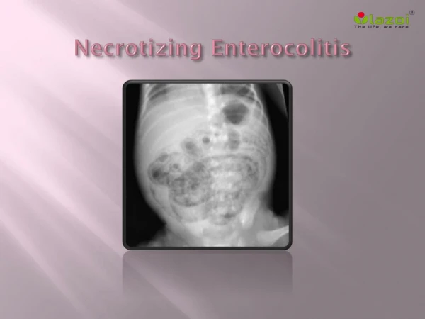
Radiographic Findings • Intestinal ileus • Dilated and thickened bowel loops, air-fluid levels • Intramural gas (pneumatosis intestinalis); cystic and/or linear patterns, terminal ileum and proximal colon • Free air (football sign) • Portal venous gas • Fixed or persistent dilated loop of bowel (sentinel loop) • Gasless abdomen with ascites
Laboratory Findings • CBC: • Elevated or decreased WBCs • Thrombocytopenia • Low ANC = poor prognosis • Elevated CRP • Cultures (blood, +/- stool, +/- CSF) • Usually reveals enteric flora • Stool Analysis - heme +, check for C. diff toxin
Laboratory Findings • Coagulopathy • Prolonged PT/PTT • Low fibrinogen • Elevated D-dimers • Electrolytes • Hypo- or hyperglycemia • Hyponatremia • Low bicarb • ABG/VBG • Metabolic acidosis
Differential Diagnosis • Sepsis with ileus • Bacterial enterocolitis: C. diff, other gram negatives • Mechanical bowel obstruction: • Hirschsprung • Ileal atresia • Volvulus • Meconium ileus • Intussusception • Isolated gastric perforation (indocin, steroids)
Gestational age (weeks) < 30 31-33 34 Full term Age at onset (days) 20 14 5 2 Mean Age at Presentation
Clinical Management Medical Vs. Surgical
Medical Management • Successfully treats ½ to 2/3 of patients • Consult surgery from the start • Bowel rest - NPO, gastric decompression, TPN • Broad spectrum antibiotics for 7-14 days • Cardiopulmonary support • Correction of metabolic acidosis and electrolyte abnormalities • Treatment of coagulopathy and/or thrombocytopenia • Serial exams, labs, and x-rays
Signs Of Ongoing Necrosis • Increasing distension • Persistent: • Metabolic acidosis • Thrombocytopenia • Hypotension from third spacing
Indications for Surgical Intervention • Severe peritonitis • Pneumoperitoneum • Intra-abdominal abscess • Positive paracentesis findings (bile & stool) • Portal venous gas seen on X-ray
Surgical Management • 34-50% of patients • Laparotomy with resection, formation of enterostomy and mucous fistula • Patch, drain, and wait • Primary peritoneal drainage • Eventual reanastomosis
Potential Complications • Short bowel syndrome • TPN-associated cholestasis with liver cirrhosis and liver failure • Catheter related sepsis • Intestinal strictures and partial small bowel obstruction • Enterocolic fistulas • Developmental and growth delay (50%)
Long-term Outcome: What’s Important? • Length of residual bowel • Ileum vs. jejunum (better adaptation) • Presence of ileocecal valve • Presence of intact colon • Maturity of infant and general condition
Patients with > 25cm of normal bowel who have an intact ileocecal valve Normal bowel length: Term infants 200-300 cm Preterm infants 100-200 cm Patients with >40cm of normal bowel who have no ileocecal valve Survival Without Transplantation
Short Bowel Syndrome • Fluid & electrolyte losses • Bile acid and Vit B12 malabsorption • Gastric acid hypersecretion inactivates pancreatic enzymes and causes fat malabsorption • Secretory diarrhea • Bacterial overgrowth - Increases malabsorption, lactic acidosis, colitis, Vit B12 deficiency
Malabsorption • Fat: Bacterial deconjugation of bile salts and acids • Protein and carbohydrates: enzyme and transport deficiencies • Vit B12: bacterial uptake
Sites of Nutrient Absorption • Duodenum: iron • Jejunum: Carbohydrates, proteins, fats and vitamins, copper • Ileum: Bile acids, Vit B12
Short Gut: Symptoms • Distension • Diarrhea • Cramping • Weight loss • Anemia (occult blood loss, Vit B12 deficiency)
Treatment of Short Gut Syndrome • Promotion of villous hyperplasia: • Drip feedings using elemental formulas • Long-chain fats stimulate intestinal adaptation • MCT diet bypasses need for bile acids • Hydrolyzed proteins absorbed rapidly • Cholestyramine (bile acid binder) • Trimethoprim-sulfa, metronidazole treats bacterial overgrowth • Proton pump inhibitors or H2 blockers
Formulas Elemental: • Require minimal digestive function and cause less pancreatic secretion • Individual amino acids or short peptides • Glucose polymers • Low fat (long chain triglycerides) • MCT absorbed in absence of lipase or bile salts
Monitoring • Stool output for fluid losses • Carbohydrate malabsorption (low stool pH or stool reducing substances) • Anticipate slow gut adaptation over years • Weight gain and growth
Prevention of NEC • Prenatal steroids • Correction of hypovolemia and hyperviscosity • Slow, gradual advancement of feeds • Breastfeeding • Probiotics - Oral immunoglobulins and bifidobacterium? • Oral antibiotics? • Acidification of feedings (avoidance of PPIs and H2 blockers)? • Glutamine or arginine supplemenation?
Trophic Feedings • No increased risk of NEC • Increases gut motility • Reduces cholestasis • Improves tolerance of subsequent feedings • May prevent gut atrophy, inflammation, and bacterial translocation