NECROTIZING ENTEROCOLITIS PRESENTED BY
NECROTIZING ENTEROCOLITIS PRESENTED BY ANSU ANN (NICU). ANTHROPOMETRIC MEASUREMENT weight: 650 gms Length: 31 cm Height: 22cm APGAR: 1/1, 6/5, 7/15

NECROTIZING ENTEROCOLITIS PRESENTED BY
E N D
Presentation Transcript
NECROTIZING ENTEROCOLITIS PRESENTED BY ANSU ANN (NICU)
ANTHROPOMETRIC MEASUREMENT • weight: 650 gms • Length: 31 cm • Height: 22cm • APGAR: 1/1, 6/5, 7/15 • Type of delivery: LSCS
HISTORY • Mother is 29y/o, G2_P1_, having uncontrolled PET, received (Labetalol, MgSO4, Ehydralazine and one dose of Dexamethasone), with gestational age of 29wks who undergone emergency CS due to PET and fetal distress.
PHYSICAL EXAMINATION • INTEGUMENTARY : skin are edematous and pinkish in color warm to touch • CVS: no heart murmur S1 S2 present, maintaining normal range of heart rate and BP, decreased peripheral perfusion • RESP: with O2 support of mechanical ventilator due to respiratory failure. • GIT: Increased abdominal girth, visible intestinal loops, abdominal distension, decreased bowel sounds, palpable abdominal mass, erythema of abdominal walls. • GUT: passing urine and stool (color of stool: dark greenish). • MS: (+) movement of extremities
INTRODUCTION • Necrotizing enterocolitis “NEC” is the most common gastrointestinal emergency in the premature infant, an important cause of neonatal morbidity and mortality. NEC affects apparently healthy premature infant who have no other medical problems or those who have recovered from their initial respiratory disease, look well and are feeding and growing. INCIDENCE • Although NEC is most commonly observed in premature infant, 10% of affected patients are born term. Between 0.3 and 2.4 infants/ 1000 birth and between 7-11% (range 3-22% in individual nursery data) among infants of less than 1500g male and female are equally affected. NEC mortality varies between 9-28
The intestines are a long, continuous tube running from the stomach to the anus. Most absorption of nutrients and water happen in the intestines. The intestines include the small intestine, large intestine, and rectum. • The small intestine (small bowel) is about 20 feet long and about an inch in diameter. Its job is to absorb most of the nutrients from what we eat and drink. Velvety tissue lines the small intestine, which is divided into the duodenum, jejunum, and ileum. • The large intestine (colon or large bowel) is about 5 feet long and about 3 inches in diameter. The colon absorbs water from wastes, creating stool. As stool enters the rectum, nerves there create the urge to defecate.
DEFINITION • NEC is the death of the intestinal tissue occurs when the lining of the intestinal wall dies and tissues falls off.
ETIOLOGY (CAUSES) • NEC occurs when the lining of the intestinal wall and tissues falls off. • Cause (unknown) • Bacteria in the intestine • Infant is already ill or premature • Prolonged hospitalization
RISK FACTORS • Premature infants • Infant who are fed by concentrated formulas • Infant who received blood exchange transfusion
SIGNS AND SYMPTOMS It may occur suddenly or slowly. Abdominal distention Feeding intolerance Blood in the stool Lethargy Diarrhea Temperature instability Vomiting Prior to any specific signs and symptoms, we can observe the activity level of the infant and temperature instability. Speed of progression of the diseases quite variable, in some cases onset is sudden little warning signs and is followed by severe apnea which require intubation, persistent metabolic acidosis, hypotension requires bolus of intravascular therapy.
PATHOPHYSIOLOGY • Infectious agent: Klebsiella, E. Coli, Clostridia, Coagulus negative Staphylococcus, • Coronavirus • EnteralElementation: NEC occurs mostly infect infants, 90%
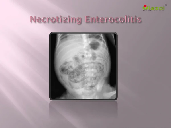
DIAGNOSIS • Radiology: The abdominal X-ray is the best diagnostic tool in the evaluation of NEC. Pneumatosisintestinalis (air the bowel wall), when present.
Laboratory evaluation: Common laboratory abnormalities include thrombocytopenia, leukocytosis, electrolytes imbalance, metabolic acidosis, hypoxia or hyper apnea; therefore one should carefully monitor the complete blood count, electrolytes and blood gases. Blood culture should be obtained before antibiotics are started. • Bell (clinical staging): • Stage 1: Suspect: Infant with suggestive clinical signs but X-ray non-diognostic. • Stage 2: Definite: Infant w/ pneumatosisintestinalis (11A: mildly ill, 11B: moderately ill (acidosis, thrombocytopenia or ascites)) • Stage 3: Advanced: (111A : critically w/ impending perforation, 111B: critical w/ proven perforation)
TREATMENT • Early bowel decompression by effective nasogastric tube suctioning. • Prompt intravenous spectrum antibiotic therapy (usually include Ampicillin, an aminoglycoside, and anaerobic bacterial coverage such as clindamycin). • Maintain volemia/ mesenteric perfusion, intravascular volume supplement is required to maintain mesenteric perfusion and to avoid worsening intestinal injury. • Except in the milder cases, because of the respiratory failure and worsening acidosis, intubation mechanical ventilation is often necessary. • Pain control is essential in this extremely painful disease, a fentanyl drip is often used at 2-4 mcg/kg/hr.
Early parenteral nutrition w/ adequate protein/ calories/ lipid is essential in order to provide substrate for the bowel to heal. • Surgical option include laparotomy w/ resection and enterostomy or peritonial drain placement, allowing abdominal decompression.
COMPLICATION • NEC complication include inadequate nutrition w/ failure to thrive, electrolytes and nutrient losses, complication due to prolonged total parenteral nutrition and central venous catheters (infections, thrombus), intestinal surgical complications (intestinal stricture in 25-35% of NEC survivors, complication related to the stoma,..) and short bowel syndrome.
PROGNOSIS • NEC mortality ranges from reported 9-28% and is due to refractory shock. • HEALTH TEACHING • Encourage all mothers to initially provide breast milk for their preterm • Educate all staff about clinical signs of NEC and increase awareness • Evaluate any untoward GI findings(abdominal distension , feeding intolerance
CONCLUSION • True reduction in the incidence of NEC likely to depend on limiting preterm • birth.Health care providers having close contact with infant play vital role in recognition and management of this potentially debilitating disease. REFERENCES • ^Sodhi C, Richardson W, Gribar S, Hackam DJ (2008). "The development of animal models for the study of necrotizing enterocolitis". Dis Model Mech 1 (2-3): 94–8. doi:10.1242/dmm.000315. PMC2562191. PMID19048070. http://dmm.biologists.org/cgi/pmidlookup?view=long&pmid=19048070. • ^Skelley, Tao Le, VikasBhushan, Nathan William. First aid for the USMLE step 2 CK (8th ed. ed.). New York: McGraw-Hill Medical. pp. 369-370. ISBN9780071761376. • ^ Marino, Bradley S.; Fine, Katie S. (1 December 2008). Blueprints Pediatrics. Lippincott Williams & Wilkins. ISBN978-0-7817-8251-7. http://books.google.com/books?id=oqpSRIOcd8MC. • ^ Hunter CJ, Upperman JS, Ford HR, Camerini V (February 2008). "Understanding the susceptibility of the premature infant to necrotizing enterocolitis (NEC)". Pediatric Research 63 (2): 117–23. doi:10.1203/PDR.0b013e31815ed64c. PMID18091350.