Human Cytogenetics
Human Cytogenetics. Dr Maha Al- Sulaimani Department of Biochemistry. What is a chromosome?. How many chromosomes do people have?. In humans, each cell normally contains 23 pairs of chromosomes, for a total of 46.

Human Cytogenetics
E N D
Presentation Transcript
Human Cytogenetics DrMaha Al-Sulaimani Department of Biochemistry
How many chromosomes do people have? • In humans, each cell normally contains 23 pairs of chromosomes, for a total of 46. • Twenty-two of these pairs, called autosomes, look the same in both males and females. • The 23rd pair, the sex chromosomes, differ between males and females. • Females have two copies of the X • chromosome, while males have one X and one Y chromosome.
How many chromosomes do people have? • The 22 autosomes are numbered by size. • The other two chromosomes, X and Y, are the sex chromosomes. • This picture of the human chromosomes lined up in pairs is called a karyotype.
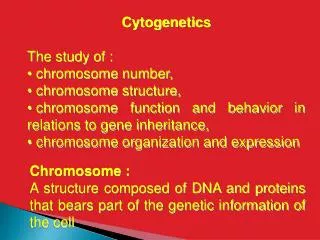
What is Cytogenetics? • Cytogenetics is the study of normal and abnormal chromosomes. • This includes examination of chromosome structure, learning and describing the relationships between chromosome structure and phenotype, and seeking out the causes of chromosomal abnormalities.
What is Cytogenetics? • In the simplest case, chromosomes are examined and characterized by obtaining an individual's karyotype, which is a description of the number and structure of the chromosomes.
Cell Cycle Phases • The cell cycle is an ordered set of events, culminating in cell growth and division into two daughter cells. • The stages are G1, S, G2, and M. • The G1 stage stands for "GAP 1". • The S stage stands for "Synthesis". • This is the stage when DNA replication occurs.
Cell Cycle Phases • The G2 stage stands for "GAP 2". • The M stage stands for "mitosis", and is when nuclear (chromosomes separate) and cytoplasmic (cytokinesis) division occur. • G0:(Preparation). • Interphase: taking in nutrients (G1, S and G2 phases). • In this stage nucleus and cytochrome division does not occur. • The cell prepares to divide.
G1 Phase • It is also called the growth phase. During this phase the biosynthetic activities of the cell, which had been considerably slowed down during M phase, resume at a high rate. • This phase is marked by synthesis of various enzymes that are required in S phase, mainly those needed for DNA replication. • Duration of G1 is highly variable, even among different cells of the same species.
S Phase • Starts when DNA synthesis commences; when it is complete, all of the chromosomes have been replicated, i.e., each chromosome has two (sister) chromatids. • Thus, during this phase, the amount of DNA in the cell has effectively doubled. • During this phase, synthesis is completed as quickly as possible due to the exposed base pairs being sensitive to external proteins such as any drugs taken or any mutagens (such as nicotine).
G2 Phase • The cell then enters the G2 Phase, which lasts until the cell enters mitosis. • Again, significant biosynthesis occurs during this phase, mainly involving the production of microtubules, which are required during the process of mitosis. • Inhibition of protein synthesis during G2 phase prevents the cell from undergoing mitosis.
M Phase (Mitosis) • Mitosis is the process by which an eucaryotic cell separates the chromosomes in its cell nucleus into two identical sets in two nuclei. • It is generally followed immediately by cytokinesis, which divides the cytoplasm, organelles and cell membrane into two cells containing roughly equal shares of these cellular components.
M Phase (Mitosis) • The relatively brief M phase consists of nuclear division (karyokinesis). • The M phase has been broken down into several distinct phases, sequentially known as: • Prophase (chromatin condensation into chromosomes), • Metaphase (alignment of chromatin in the middle), • Anaphase (chromosomes move to opposite poles), • Telophase (two daughter nuclei form in the cell), • Cytokinesis (strictly speaking, cytokinesis is not part of mitosis but is an event that directly follows mitosis in which cytoplasm is divided into two daughter cells).
M Phase (Mitosis) • Mitosis and cytokinesis together define the mitotic (M) phase of the cell cycle: the division of the mother cell into two daughter cells, genetically identical to each other and to their parent cell. • This accounts for approximately 10% of the cell cycle.
Control of the Cell Cycle • How cell division (and thus tissue growth) is controlled is very complex. • The following terms are some of the features that are important in regulation, and places where errors can lead to cancer.
Control of the Cell Cycle • Cdk (cyclin dependent kinase, adds phosphate to a protein), along with cyclins, are major control switches for the cell cycle, causing the cell to move from G1 to S or G2 to M. • MPF (Maturation Promoting Factor) includes the CdK and cyclins that triggers progression through the cell cycle. MPF is activated at the end of G2 by a phosphatase, which removes an inhibitory phosphate group added earlier (by CDK).
p53 is a protein that functions to block the cell cycle if the DNA is damaged. If the damage is severe this protein can cause apoptosis (cell death). • p53 levels are increased in damaged cells. This allows time to repair DNA by blocking the cell cycle. • A p53 mutation is the most frequent mutation leading to cancer. An extreme case of this is Li-Fraumeni syndrome, where a genetic a defect in p53 leads to a high frequency of cancer (breast and bone) in affected individuals.
Control of the Cell Cycle • p27 is a protein that binds to cyclin and cdk blocking entry into S phase. Recent research (Nature Medicine 3, 152 (1997)) suggests that breast cancer prognosis is determined by p27 levels. • Reduced levels of p27 predict a poor outcome for breast cancer patients.
Basics of Meiosis • Meiosis: Two consecutive cell divisions. • Creation of gametes or sex cells. • Each subsequent daughter cell is haploid (n) as opposed to diploid(2n) like in mitosis.
Meiosis 1 • Prophase 1 • Metaphase 1 • Anaphase 1 • Telophase 1
Spindle fibers Nucleus Nuclear envelope Prophase I (early) (diploid) Prophase I (late) (diploid) Metaphase I (diploid) Anaphase I (diploid) Telophase I (diploid) Meiosis 1
Meiosis II : The Equational Division • Prophase 2 • Metaphase 2 • Anaphase 2 • Telophase 2
Results of Meiosis • Gametes: • Female = ovum(egg) • Male = sperm • Four haploid cells. • One copy of each chromosome. • One allele of each gene. • Different combinations of alleles for different genes along the chromosome.
Animations • Gametogenesis: Flash animation for sperm creation: http://highered.mcgraw-hill.com/olc/dl/120112/anim0043.swf • Oogenesis: Flash animation for ovum creation: http://msjensen.education.umn.edu/1135/Links/Animations/Flash/0044-swf_maturation_of_.swf
Haploid Ovum/Egg Haploid Ovum/Egg