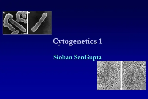
Cytogenetics 1 Sioban SenGupta
510 likes | 1.14k Vues
Definitions. CytogeneticsVisual study of chromosomes at microscopic levelKaryotypeChromosome complement ? also applied to picture of chromosomesIdiogramStylised form of karyotype. Chromosomal abnormalities. 1959 - Down syndrome (LeJeune)1970 - banding techniquesidentification of individual

Cytogenetics 1 Sioban SenGupta
E N D
Presentation Transcript
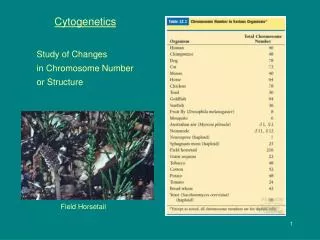
2. Definitions Cytogenetics
Visual study of chromosomes at microscopic level
Karyotype
Chromosome complement
� also applied to picture of chromosomes
Idiogram
Stylised form of karyotype
3. Chromosomal abnormalities 1959 - Down syndrome (LeJeune)
1970 - banding techniques
identification of individual chromosomes
Karyotype and FISH
types of abnormalities:
Extra copy of chromosome
Missing copy of chromosome
Structural abnormalities
4. Chromosomes Centromere - movement during cell division
divides the chromosomes into short (p) and long (q) arms
Telomere - tip of each chromosome
seal chromosomes and retain chromosome integrity
telomere consists of tandem repeats TTAAGGG
maintained by enzyme - telomerase
reduction in telomerase and decrease in number repeats important in ageing and cell death
5. Chromosomes Classified according to position of centromere
Central centromere - metacentric
Sub-terminal centromere - acrocentric
have satellites which contain multiple copies of genes for ribosomal RNA
Intermediate centromere - submetacentric
6. Chromosomes
7. Chromosomes 22 autosomes and sex chromosomes in pairs
Classified according to:
Length
position of centromere
presence or absence of satellites
Chromosomes divided into groups labelled A-G
8. Karyotyping Staining methods to identify chromosomes
G banding - Giemsa
Q banding - Quinacrine
R banding - Reverse
C banding - Centromeric (heterochromatin)
Ag-NOR stain - Nucleolar Organizing Regions (active)
9. Karyotyping � cell preparation Need metaphases
Culture cells until sufficient mitotic activity
Add colchicine (or colcemid) to arrest in metaphase
prevents mitotic spindle fibres forming
Add hypotonic salt solution to swell cells
Fix with mix of methanol;acetic acid
Want long chromosomes with none overlapping
10. G banding Most common method used
Chromosomes treated with trypsin
denatures protein
Giemsa stain
each chromosome characteristic light and dark bands
400 bands per haploid genome
Each band corresponds to 5-10 megabases
High resolution (800 bands ; prometaphase chromosome)
use methotrexate and colchicine
Dark bands are gene poor
11. Preparation of G banded karyotype
12. G banding Metaphase spreads
Count chromosomes in 10-15 metaphases
If mosaicism suspected, count 30
Detailed analysis of 3-5 metaphases
Used to photograph and cut out
Now computer programmes
13. Normal male karyotype
14. Normal female karyotype
15. Q banding Used especially for Y chromosome abnormalities or mosaicism
Similar pattern to G banding
But can detect polymorphisms
Needs fluorescent microscope
16. R banding Used to identify X chromosome abnormalities
Heat chromosomes before staining with Giemsa
Light and dark bands
are reversed
17. C banding Used to identify centromeres / heterochromatin
Heterochromatic regions
contain repetitive sequences
highly condensed chromatin fibres
Treat with chromosomes with
Acid
Alkali
Then G band
18. Idiogram
19. ISCN International System for Human Cytogenetic Nomenclature
Each area of chromosome given number
Lowest number closest (proximal) to centromere
Highest number at tips (distal) to centromere
20. ISCN del - deletion
dic - dicentric
fra - fragile site
i - isochromosome
inv - inversion
p - short arm
r - ring
der - derivative
dup - duplication
h - heterochromatin
ins - insertion
mat - maternal origin
q - long arm
t - translocation
21. ISCN , 46,XX,del(5p)
separates
chromosome numbers
sex chromosomes
chromosome abnormalities
; 46,XX,t(2;4)(q21;q21)
separates
altered chromosomes
break points in structural rearrangements involving more than 1 chromosome
22. ISCN Normal male
46,XY
Normal female
46,XX
23. Types of chromosome abnormalities Numerical
Aneuploidy (monosomy, trisomy, tetrasomy)
Polyploidy (triploidy, tetraploidy)
Structural
Translocations
Inversions
Insertions
Deletions
Rings
Isochromosomes
ESAC
24. Numerical Aneuploidy
Autosomal trisomy, 47
Sex chromosomes, 45, 47, 48, 49
Polyploidy
Whole chromosome set
Triploidy, 69
Tetraploidy, 92
25. Aneuploidy Almost all been found in oocytes and early embryos, trisomies and monosomies
Most lethal (miscarry)
Do not see in pregnancy or live born
Exceptions sex chromosomes and Down
Some aneuploidy is age related
26. Sex chromosomes Abnormalities more tolerated
If have extra Y, few genes mainly for sex determination
If have extra X, excess X is inactivated
Monosomy X, Turners
Majority die during development
Only small proportion survive to birth
Short and infertile
27. Sex chromosome abnormalities Turner Syndrome 45,XO (female)
Trisomy X��������� 47,�XXX (female)
Klinefelter Syndrome 47,XXY (male)
Extra �Y� chromosome 47,XYY (male)
28. Down syndrome, trisomy 21
29. Edwards syndrome, trisomy 18
30. Patau syndrome, trisomy 13
31. Structural Breakage in at least 1 chromosome
Translocations
2 different chromosomes break and rejoin incorrectly
Inversions
2 breaks in same chromosome
Insertions
Piece of chromosome inserted
Deletions
Piece of chromosome missing
32. Chromosome breaks Once chromosome broken by some means
Unstable situation as telomeres not at end
Usually join up to other piece
33. Translocations Chromosome moves from normal position
to abnormal position
Robertsonian
Acrocentric chromosomes
D and G groups
(13, 14, 15, 21, 22)
Reciprocal
Any chromosome
34. Robertsonian translocations Lose satellite and short arms
Genes for rRNA
Repeated on other acrocentric chromosomes
Reduce chromosome number by one (45)
but no loss of chromatin from long arms
Phenotypically normal � problems at meiosis
Involved in evolution
35. Robertsonian translocations
36. Robertsonian translocations D:G translocation
Often 14:21 joined
G:G translocation
21:22 joined
21:21 joined
21 smallest chromosome
37. Robertsonian translocation family pedigree
38. Robertsonian translocations
45,XY,der(13q;14q)(q10;q10)
45,XX,der(13q;21q)(q10;q10)
39. Reciprocal translocations More common than Robertsonian
Break in any chromosome at any point
Phenotypically normal � problems at meiosis
40. Reciprocal translocation 46,XX,t(5;9)(q32;p13)
41. Inversions Reversal of segment of chromosome
If too small cannot detect by karyotype
Very rare in humans
Selected against as would get reduced fertility
Pericentric
reversed segment includes centromere
Paracentric
within one chromosome arm
Paracentric inversion
main difference in karyotypes of great apes and humans so important in evolution
42. Inversions
Pericentric Paracentric
43. Insertions Segment of 1 chromosome inserted into another
44. Deletions Terminal
loss of end of chromosome
46,XY,del(10)(q26) missing long arm of 10
Interstitial
loss of segment from within chromosome
46,XY,del(10)(q24q26) missing segment of 10
All result in unbalanced karyotype
Partial monosomy
Serious clinical effect
45. Ring chromosome
46. Isochromosome Two copies of the same arm
Mirror image around centromere
Centromeres part in wrong plane
Monosomy for 1 chromosome arm
Trisomy for the other arm
47. ESAC Extra Structurally Abnormal Chromosome
Abnormal chromosome in addition to 46
Small and difficult to identify
Sometimes called marker chromosomes
Difficult to work out effect on person
May be benign or cause serious mental handicap