Download
1 / 25
260 likes | 462 Vues
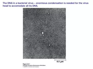
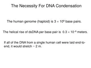
The Necessity For DNA Condensation. The human genome (haploid) is 3 10 9 base pairs. The helical rise of dsDNA per base pair is 0.3 10 –9 meters. If all of the DNA from a single human cell were laid end-to-end, it would stretch ~ 2 m. Chromatin Structure and Properties.

E N D
The Necessity For DNA Condensation The human genome (haploid) is 3 109 base pairs. The helical rise of dsDNA per base pair is 0.3 10–9 meters. If all of the DNA from a single human cell were laid end-to-end, it would stretch ~ 2 m.
Chromatin Structure and Properties 1. Structure of the histone, the nucleosome, and higher-order chromatin structure 2. Roles of nucleosomes in repressing transcription 3. Local and global methods to identify nucleosome positions 4. Transcription regulation by histone acetylation
SDS-PAGE of Histone Proteins • Small and highly basic • Highly abundant • Highly conserved • All except H1 in core nucleosome 20 kDa 15 kDa 10 kDa
Electron Micrograph of Nucleosomes (1975) • Pierre Chambon and co-workers • Removed histone H1 with trypsin or high salt concentration • Named the bead-like structures nucleosomes 500 nm
Structure of the Nucleosome (1991) H3/H4 H2A/H2B • Evangelos Moudrianakis and co-workers • x-ray crystallography provided moderate resolution structure of the histone core (3.1 Å) • suggested a possible path for DNA
Structure of the Nucleosome (1997) • Timothy Richmond and co-workers • First nucleosome structure with DNA • 147 base pairs visible along with histone octamer (H1 was not present)
Fig. 13.6 EM: 30 nm Fiber of Chromatin • Panels A-C: Low ionic strength Beads on a string • Panel D: Moderate ionic strength • Panels E-G: Higher ionic strength Formation of 30 nm fiber (100 mM NaCl) 100 nM
Fig. 13.7 Structure of a Tetranucleosome (2005) • Again, Richmond and co-workers • Structure is only 9 Å resolution, but model constructed from higher resolution structures of single nucleosomes
Structural Model of the 30 nm Fiber • Idealized structure from arranging tetranucleosomes so that angles between units are constant and steric overlap is avoided • Structure leaves unclear the role of histone H1
Model For Higher-order Chromatin Structure Nuclease studies suggested ‘circular’ DNA, even for eukaryotes Estimates for loop size range from 35 kb to 83 kb
Inactive Chromatin Lacks H1 (1984) 0.6 M KCl Certain 5S rRNA genes are transcriptionally active in Xenopus oocytes but inactive in somatic cells (Transcription is by RNA polymerase III) A. Histones in accidentally de-repressed chromatin B. Histones in purposely de-repressed chromatin, treated to remove histone H1
Competition Between Histones and Transcription Factors Both complexes are apparently long-lived, so whichever complex forms first ‘wins’ for controlling transcription
High Concentrations of Core Histones Can Also Repress Transcription (1991) • James Kadonaga and co-workers • Here, RNA polymerase II transcription followed, of a Drosophila gene • Physiological core histone level gave 4-fold repression (lane 5)
Competing Effects of Activators and Histones • Addition of H1 in addition to core histones further decreased transcription (odd numbered lanes) • Activator GAL4-VP16 had larger effect in the presence of H1, effectively reversing H1 effect
A Model of Transcriptional Activation Histone H1 Core histones
Genomic Methods of Nucleosome Mapping Oliver Rando and co-workers, 2005 Microarrays used to determine positions of nucleosome throughout yeast genome Most nucleosomes in yeast are well-positioned Linker CHA1 promoter region Positioned nucleosome Delocalized nucleosome
Promoter Regions Are Nucleosome-depleted Depleted regions
Nucleosome Position Preferences Are Present In vitro Eran Segal, Jonathan Widom and co-workers, 2008 Yeast genomic DNA isolated and reassembled with purified chicken histones Positions of histones largely match those determined in vivo using same method Suggests that nucleosome positions are globally determined by DNA sequence
+ H H An Additional Layer of Complexity: Histones Are Modified by Acetylation • Histone acetylation activates transcription, reducing the ability of nucleosomes to repress • Nuclear acetylation of core histone N-terminal tails • (histone acetyltransferase, HAT) • Catalyzed by HAT A (isolated in 1996) • Acetylate lysines within tails of core histones • Some HAT As are also transcriptional activators, suggesting that they bind near transcription start site and acetylate nearby histones
An Additional Layer of Complexity: Histones Are Modified by Acetylation • Histone acetylation activates transcription, reducing the ability of nucleosomes to repress • Nuclear acetylation of core histone N-terminal tails • (histone acetyltransferase, HAT) • Catalyzed by HAT A (isolated in 1996) • Acetylate lysines within tails of core histones • Some HAT As are also transcriptional activators, suggesting that they bind near transcription start site and acetylate nearby histones H3C
An Additional Layer of Complexity: Histones Are Modified by Acetylation • Histone acetylation activates transcription, reducing the ability of nucleosomes to repress • Nuclear acetylation of core histone N-terminal tails • (histone acetyltransferase, HAT) • Catalyzed by HAT A (isolated in 1996) • Acetylate lysines within tails of core histones • Some HAT As are also transcriptional activators, suggesting that they bind near transcription start site and acetylate nearby histones • Cytoplasmic acetylation carried out by HAT B • Prepares histones for incorporation into nucleosomes • Acetyl groups later removed in nucleus
Model For Repression By Histone Deacetylase Retinoic acid receptor heterodimer
Histone Acetylation • How does acetylation increase transcription? • Reduces positive charge on lysine-rich regions, presumably reducing affinity of nucleosome for DNA • May weaken or disrupt interactions between nucleosomes • Can recruit transcription factors (bromodomain)
1. Cellular DNA is packaged with histone proteins into a higher order structure called chromatin. The first level of chromatin organization is the nucleosome, and a higher order structure is the 30 nm fiber. Key Points 2. The packaging, or condensation, of DNA into chromatin represses transcription because the transcription machinery cannot efficiently access the promoter. 3. Nucleosomes are globally depleted near transcription start sites, and their positions appear to be greatly influenced by DNA sequences. 4. Modification of histones by acetylation activates transcription.