Methods
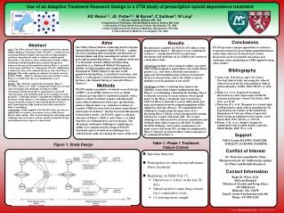
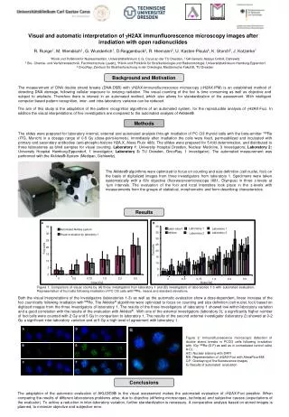
A. 35. Automated Aklides system. 30. Visual evaluation by laboratory 1. 25. 20. Foci/cells. 15. 10. 5. 0. 0. 0,5. 0,75. 1,0. 2,0. 5,0. Dose [Gy]. B. 40. Laboratory 1. Labor atory1. Laboratory 1. 35. Aklides. Laboratory 3. Laboratory 2. 30. 25. Foci / cells. 20. 15.

Methods
E N D
Presentation Transcript
A 35 Automated Aklides system 30 Visual evaluationbylaboratory 1 25 20 Foci/cells 15 10 5 0 0 0,5 0,75 1,0 2,0 5,0 Dose [Gy] B 40 Laboratory 1 Labor atory1 Laboratory 1 35 Aklides Laboratory 3 Laboratory 2 30 25 Foci/cells 20 15 10 5 0 0 0,5 0,75 1,0 2,0 5,0 Dose [Gy] R. Runge1, M. Wendisch1, G. Wunderlich1, D.Roggenbuck2, R. Hiemann3, U. Kasten-Pisula4, K. Storch5, J. Kotzerke11Klinik und Poliklinik für Nuklearmedizin, Universitätsklinikum C.G. Carus an der TU Dresden, 2 GA Generic Assays GmbH, Dahlewitz3Bio- Chemie- und Verfahrenstechnik, Fachhochschule Lausitz, 4Klinik und Poliklinik für Strahlenbiologie und Radioonkologie, Universitätsklinikum-Hamburg-Eppendorf5 OncoRay, Zentrum für Strahlenforschung in der Onkologie, Medizinische Fakultät, TU Dresden Visual and automaticinterpretationofgH2AX immunfluorescencemicroscopyimages after irradiationwith open radionuclides Background and Motivation The measurementof DNA double strandbreaks (DNA-DSB) withgH2AX-immunofluorescence microscopy (gH2AX-IFM) is an establishedmethodofdetecting DNA damage, followingcellularexposuretoionizingradiation. The visualcountingofthefociis time consumingas well asobjective and subjecttoartefacts. Thereforethereisinterest in an automatedmethod, which also allowsforstandardizationoftheassessment. With intelligent computerbasedpatternrecognition, inter- and intra-laboratoryvariancecanbereduced. The aimofthisstudyistheadaptationofthepatternrecognitionalgorithmsof an automatedsystem, forthereproducibleanalysisofgH2AX-Foci. In additionthevisualinterpretationsoffiveinvestigatorsarecomparedtotheautomatedanalysisofAklides®. Methods The slideswerepreparedforlaboratoryinternal, external and automatedanalysisthroughirradiationof PC Cl3 thyroidcellswiththebeta-emitter188Re (ITG, Munich) in a dosagerangeof0-5 Gy(dose-point-kernels). Immidiately after irradiationthecellswerefixed, permeabilized and incubatedwithprimary and secondaryantibodies (anti-phospho-histoneH2A.X, Alexa Fluor 488). The slildeswerepreparedfor 5-fold determination, and distributedtothreelaboratoriesas blind samplesforvisualcounting. Laboratory 1: University Hospital Dresden, NuclearMedicine, 3 investigators; Laboratory 2:University Hospital Hamburg-Eppendorf, 1 investigator; Laboratory 3: TU Dresden, OncoRay, 1 investigator). The automatedmeasurement was performedwiththeAklides®-System (Medipan, Dahlewitz). The Aklides® algorithmswereoptimizedtofocus on counting and sizedefinition (cellnuclei, foci) on thebasisofdigitalizedimagesfromthreeinvestigatorsfromlaboratory 1. Specimensweretakensystematicallywith a 60x objective (fluorescencemicroscopeIX81, Olympus) in three z-levelsat 1µm intervals. The evaluationofthefoci and localintensitiestookplace in the z-levelswithmeasurementsfromthegroupsofstatistical, morphometric and form-describingcharacteristics. C Results Figure1: Comparisonofvisualcountsby (A) threeinvestigatorsfromlaboratory 1 and (B) investigatorsoflaboratories 1-3 withautomatedevaluation. Representationoffoci/cellsfollowingirradiationof PC Cl3 cells with188Re, means and standarddeviations. Boththevisualinterpretationsoftheinvestigators (laboratories 1-3) as well astheautomaticevaluationshow a dose-dependent, linear increaseofthefocicount/cellsfollowingirradiation with188Re. The Aklides®algorithmswereoptimizedtofocus on counting and sizedefinition (cellnuclei, foci) based on digitizedimagesfromthethreeinvestigatorsoflaboratory 1. The resultsofthethreeinvestigatorsoflaboratory 1 showedlowwithin-laboratoryvariation and a goodcorrelationwiththeresultsoftheevaluationwithAklides®. Withoneoftheexternalinvestigators (laboratory 3), a significantlyhighernumberoffoci/cellswerecountedwith 2 Gy und 5 Gyin comparisontolaboratory 1. The resultsofthesecondexternalinvestigator (laboratory2) showedat 0-2 Gya significant inter-laboratoryvariation and at 5 Gya highlevelofagreementwithlaboratory1. Figure 2: Immunofluorescence microscopicdetectionof double strandbreaks in PCCl3 cellsfollowingirradiationwith 1Gy 188Re (D-F) as well as in unirradiatedcontrolcells( A-C). A/D: Nuclearstainingwith DAPI B/E: RepresentationofgH2AX-Foci with AlexaFluor488C/F: Overlayingofthefluorescenceimages G: Resultsofautomatedevaluation Conclusions The adaptationoftheautomaticevaluationof AKLIDES® tothevisualassessmentmakestheautomatedevaluationofgH2AX-Foci possible. Whencomparingtheresultsof different laboratoriesproblemsarise, due toobjective (differingmicroscopes, technique) and subjectivecauses (expectationsoftheevaluator). Toachive a reduction in inter-laboratoryvariation, furtherstandardizationisnecessary. A comparativeanalysisbased on storedimagesisplanned, tominimizeobjective and subjectiveerror.