P lacode ectoderm and the neural crest : d evelopment and derivatives Dr. Altdorfer
P lacode ectoderm and the neural crest : d evelopment and derivatives Dr. Altdorfer. 3 week-old human embryo. Placodes - and neural crest. Placode : cranial , lateral , epithelialial tickenings : „ Plaque ” may form vesicles.

P lacode ectoderm and the neural crest : d evelopment and derivatives Dr. Altdorfer
E N D
Presentation Transcript
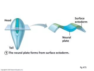
Placode ectoderm and the neural crest: development and derivatives Dr. Altdorfer
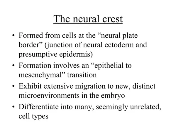
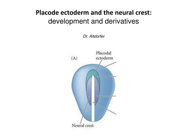
Placodes • - and neuralcrest Placode:cranial, lateral, epithelialialtickenings: „Plaque” mayformvesicles Neuralcrest:caudal, medial. EMT: epithelio-mesenchymaltransition Prof. Szél
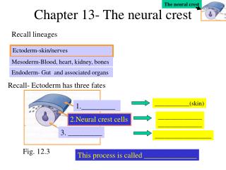
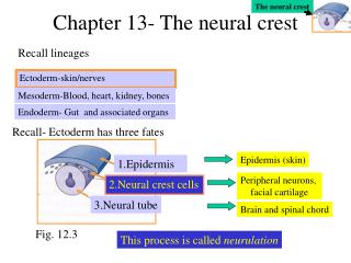
Placode Derivative Hypophyseal Olfactory Lens Trigeminal Otic Epibranchial Neural plate Neural crest Ectoderm Rathke’s pouch Adenohypophysis Olfactory epithelium Lens Trigeminal ggl. (partly) Otic vesicle membranous labyrinth, spiral+vestibular ggl.* (VIII.) Epibranchial –> geniculate ggl. (VII.), inf. ggl. of IX. and X. nerves* Brain, spinal cord ganglia, … epidermis of skin,… *Special sensory ganglia! after Webb and Noden (1993)
2. Neuralcrest 3 week-old human embryo
Ectoderm/neuroectoderm specification • Molecularregulation of NC development, NCinduction • The margin of theneuralplate and theneighbouringectoderm, fromwhichepidermisdevelops, influenceeachother. • The concentration of BMP4 and BMP7 is • high - inthefutureepidermis, • low - intheneuralplate(because here chordin and follistatindecreasetheirexpression). • medium - intheareas of thefutureneurelcrest. snail-1 and slug (snail-2) expression starts which is needed for EMT(epithelio-mesenchymal transition) slug is expressed at gastrulation as well! Changes in adhesion properties (adhesion molecules) C. Milet, A.H. Monsoro-Burq / Developmental Biology 366 (2012) 22–33
After induction: migration ECM: Fibronectin, laminin, and type IV. collagen are favorable ECM: chondroitin-sulfate: non-favorable cell-surface integrins TRUNK HEAD In the head region neural crest cells start the migration before the closure of the neural tube, not in the trunk
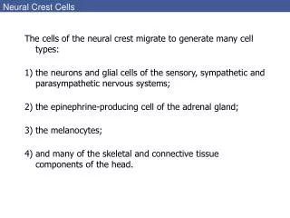
Neural crest - derivatives Neural crest cells are a multipotent progenitor population, so that the fates of specific crest populations must be controlled through environmental factors during normal development. http://www.embryology.ch
Neural crest cells are a multipotent progenitor population, the fates of specific crest populations must be controlled through environmental factors during normal development. In the trunk region the neural crest cells are divided into two groups: - Those which migrate dorsally, differentiate to melanocytes in the skin and hair follicles. - migrating through the ventral pathway can form sensory ganglia and the accompanying satellite cells and Schwann cells, or form autonomic ganglia: sympathetic and enteric neurons, satellite cells, Schwann cells, and cells of the adrenal medulla. • The neural crest cells of the head region have more opportunities: • Neural derivatives: • - Sensory ganglia (general sensory ones): • -superior ganglia of the IX. and X nerves, • -part of the trigeminal ganglion(other part: from placode). • Autonomic ganglia: ciliary (III.), pterygopalatine, submandibular (VII.) • and otic (IX.) ganglia (all these from 2nd rhombomer – located next to CN V. branches) • “ectomesenchymal” tissue: • -bones of the skull (frontal, parietal bones, squama of the temporal bone, nasal, palatine bones, vomer, maxilla and mandible) • -all meninges , choroid and sclera of the eye • -dentin of the teeth • -connective tissue of the glands (lacrimal, nasal, labial, palatine, oral), thyroid and parathyroid glands and thymus • -connective tissue of the head (including melanocytes), cartilages, ligaments, and tendons • -tunica media of the outflow tract of the heart and the great vessels (conotruncal septum)
Migratory pathways Trunk neural crest
Neural crest cranial NC migratingintotheskull and branchialarches; cranialmesenchyme, cranialgangliapia, arachnoidea cardiac NC - aorticopulmonaryseptum (intruncus arteriosus) vagal NC -entericnervoussystem(intramurale ggl.) in GI tract (frompharynxtill colon)-parafollikular (C-) cellsinthyroidgland • trunk NC DRG (nervecells + supportingcells)sympathetictrunkadrenalmedulla • lumbosacral NC intramural ggl. (colon)
The circumpharyngeal neural crest arises in the posterior rhombencephalic region, and in the lower part of the pharynx, emigrating circumpharyngeal crest cells pass behind the sixth pharyngeal arch. Neural crest cells from the anterior rhombencephalon to the level of somite 5 emigrate from the circumpharyngeal crest as a stream, called the cardiac crest, toward the developing heart and aortic arches, whereas other neural crest cells from the levels of somites 1 to 7 constitute the vagal crest and migrate into the developing gut as precursors of the parasympathetic innervation of the digestive tract.
19-day old chicken embryo colon Remak ganglion plexus myentericus plexus submucosus Dr. Nagy
Hirschprung’s disease (megacolon congenitum aganglionare) -developmental anomaly; affects 1:5000 human infants. • complete absence of ENS in the distal bowel (no relaxation!); proximal colon becomesdistended • 90% are diagnosed as newborns. • Failure to pass stool within 1st 2 days of life, abdomen distended, vomiting Dr. Nagy
Head neural crest • NC cell migration in 3 streams: • ‘trigeminal’–around 3 divisions of CN V. (1st branchial arch*+frontonasal process) • ‘hyoid’– into 2. branchial arch • ‘postotic’ (= behind otic vesicle) – into branchial arches 3-6. • Ectomesenchym: bones, cartilage, conn. tissue, vessels • * 2 of 3 auditory ossicles, jaw (Meckel’s cartilage)- skull… „new head” Rhombomeres 1,2,(3) Rhombomeres (3),4,(5) Rhombomeres (5),6,7,8
Head neural crest • NC cell migration in 3 streams: • ‘trigeminal’–around 3 divisions of CN V. (1st branchial arch*+frontonasal process) • ‘hyoid’– into 2. branchial arch • ‘postotic’ (= behind otic vesicle) – into branchial arches 3-6. • Ectomesenchym: bones, cartilage, conn. tissue, vessels • * 2 of 3 auditory ossicles, jaw (Meckel’s cartilage)- skull… „new head” Rhombomeres 1,2,(3) Rhombomeres (3),4,(5) Rhombomeres (5),6,7,8
Head neural crest Tooth development
Head neural crest NC participation in the ganglia
New Head Hypothesis Plasticity of neural crest cells… ecto-mesenchyme Specialcomposite sense organs, complex visual organ is important in predation. Jawsfor predation! NC Pigment cells against UV radiation, accomodation to environment. Complex viscerocranium andchondrocraniumdevelopment. • Elements of cranial organization at the core of the `New Head Hypothesis'. The New Head Hypothesis (NHH) of Carl Gans and Glenn Nothcutt (Gans and Northcutt, 1983), as schematized in mouse (A-D) and human (E) embryos. • Schematics of embryonic day 9.5 mouse embryos, highlighting specific major tissue components of the NHH. • Vertebrates exhibit unique characteristics, such as • (A) an expanded alar plate of the forebrain; • (B) formation of neurogenic sensory placodes from the surface cephalic ectoderm; • (C) a migratory mesenchymal population of cranial neural crest cells that invade the branchial arches (BA) and surround the expanded forebrain and rostral primary sensory placodes; and • (D) an expanded skeletal system to support the new rostrally expanded forebrain, rostral sensory capsules and BAs. • (E) The hypothesized shift from a passive suction feeding protovertebrate to a predatory vertebrate with jaws made from modified BA elements necessitated a high degree of craniogenic developmental and functional integration. • Abbreviations: BA, branchial arches; di, diencephalon; hb, hindbrain; mb, midbrain; olf, olfactory; op, optic; ot, otic; tel, telencephalon On the trail of the `new head' in Les Treilles Marianne Bronner-Fraser, Development 2008 135: 2995-2999; doi: 10.1242/dev.019901
DiGeorge syndrome - hoxa-3 gene defect Mesenchymalelements of cranialiscrest (III-IV. pharyngealarches) aredefective Aplasia of thymus and parathyroidgland(III-IV. pharyngealpouches), „fish-mouth” deformation (shorterphiltrum), deformation of lingual and cervicalmuscles, hypertelorism, malformations of theheart Prof. Szél
References Schoenwolf, Bleyl, Brauer, Francis-West: Larsen’s Human Embryology, Elsevier S. F. Gilbert: Developmental Biology, Sinauer associates, Inc. Publishers T. W. Sadler: Langman’s Medical Embryology, Williams & Wilkins B. M. Carlson: Human Embyology and Developmental Biology Lectures of Anatomy Department (Prof. Szél Á., Prof. Csillag A., Prof. Kálmán M. Dr. Nagy N., Dr. Kocsis K., Dr. H.-Minkó K.) Neural regulation of human life processes – from the neuron to the behaviour. Interdisciplinary teaching material concerning the structure, function and clinical aspects of the nervous system for students of medicine, health and life sciences in Hungary University of Pecs; Dialóg Campus Publishing-Nordex Kft.