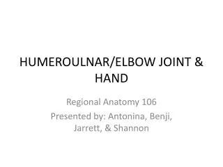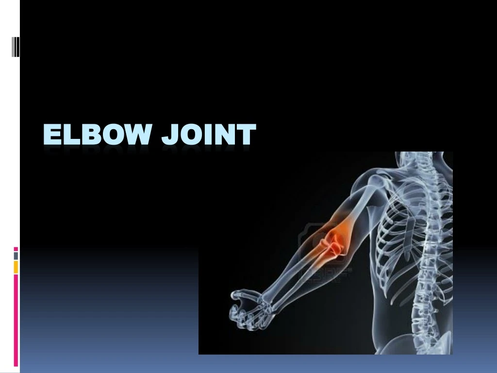

Elbow joint
E N D
Presentation Transcript
Elbow joint - Classification • Compound – more than two bones are involved • Type: Synovial • Subtype: Hinge joint • Movements – allows flexion & extension • Uniaxial joint
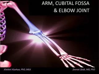
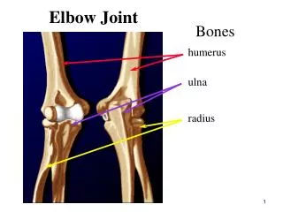
Elbow Joint Three articulations • Humerus and Ulna • Humerus and radius • Radius and ulna --cubital articulation
Elbow joint – Carrying angle • Angle between the arm and forearm • 163o • To prevent the ulnar border of the forearm from rubbing against the hip • Facilitates carrying of bulky objects
Elbow joint – Carrying angle Cubitusvalgus • Deformity of the elbow in which it deviates away from the midline of the body when extended. • One of the clinical feature of Turner syndrome(45, XO) Cubitusvarus • Deformity of the elbow in which it deviates toward the midline of the body when extended. Cubitus valgus Cubitus varus
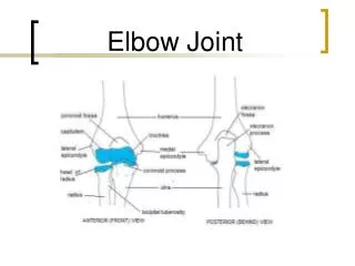
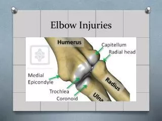
Elbow joint - Articular surfaces Olecranon fossa Radailfossa Olecranon process Coronoidfossa capitulum Trochlea Head of radius Radial notch of ulna
Elbow joint - Ligaments • Capsule • Envelops the joint completely • Thin enough to allow for movements • Strengthened by collateral ligaments • Attached to the margins of articular surfaces • Lined by synovial membrane on the inner aspect
Elbow joint – Nerve supply • Musculocutaneous • Radial • Ulnar
FRACTURES OF ELBOW • DISTAL HUMERUS: FRACTURES: • SUPRACONDYLAR • DISTAL HUMERUS: FRACTURES: • SUPRACONDYLAR • INTERCONDYLAR • CONDYLAR • EPICONDYLAR • FRACTURES OF PROXIMAL ULNA: • OLECRANON FRACTURE • CORONOID PROCESS FRACTURE • FRACTURES OF RADIUS: • RADIAL HEAD • RADIAL NECK
SUPRACONDYLAR FRACTURE • This is the most common fracture to occur around the elbow in children (60%). • Fragment gets displaced anteriorly • .
The fracture line extends transversely or obliquely through the distal humerus above the condyles.
Medial epicondylitis GOLFER’S ELBOW • repetitive motions with poor technique, muscular weakness or anatomical weakness • often seen in the acceleration phase of throwing