Shoulder Trauma
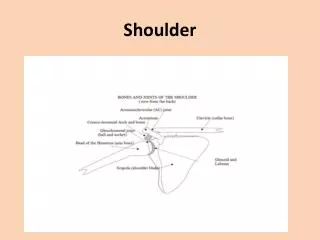
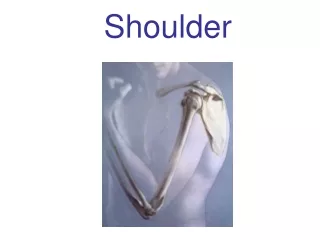
Shoulder Trauma. Normal anatomy. Standard AP shoulder series demonstrates most of the essential anatomy Internal rotation, external rotation, abduction (baby arm) Specialized views may be required to reduce overlap of certain structures

Shoulder Trauma
E N D
Presentation Transcript
Normal anatomy • Standard AP shoulder series demonstrates most of the essential anatomy • Internal rotation, external rotation, abduction (baby arm) • Specialized views may be required to reduce overlap of certain structures • http://eorif.com/Shoulderarm/XrayShoulder.html http://www.ski-injury.com/specific-injuries/shoulder
Clavicle Fractures • 15% of all fractures; most common fracture during birth • Usually direct trauma • Males 2/3 of all clavicle fractures • More common in children and adolescents; incidence decreases with age
Clavicle fractures by location • Medial 1/3- least common (5%) • Middle 1/3- most common (75%) • If fracture is complete, medial fragment will be elevated by action of SCM and lateral fragment will be depressed by the weight of the upper extremity • Distal 1/3- (20%) • Fracture may extend and become intra-articular http://eorif.com/Shoulderarm/Clavicle%20medial.html
Clavicle fracture complications • Child and adolescent heal without complication 95% of the time • Increased chance of complications in adults • Complications include: • Neurovascular damage • Non-union • Mal-union • Degenerative arthritis • Post-traumatic osteolysis
Neurovascular damage • Most commonly subclavian artery; less commonly subclavian vein; occasionally brachial plexus and sympathetic chain http://bestpractice.bmj.com/best-practice/monograph/592/basics/aetiology.html
Non-union • 5% of cases • Lack of callous formation by 6 weeks post-injury signifies non-union • Radiographic signs: • Fracture margins become sclerotic and rounded with smooth contours over time • May require surgery http://www.gentili.net/fracture.asp?ID=16
Malunion • If bones overlap and massive callus develops, cosmetic deformity and functional impairment results • May require surgery http://www.sciencedirect.com/science/article/pii/S1058274604002678
Degenerative Arthritis • Painful arthritis follows untreated intra-articular fractures • Radiographic signs are the usual findings in OA http://www.drmaffet.com/shoulder-surgery-houston/ac-joint-arthosis-2/
Post-traumatic Osteolysis • Bone resorption of distal clavicle • First becomes radiographically visible 2- 3 months after the injury • Distal cortex becomes hard to define and may become tapered over time • Injury may be trivial, not necessarily fracture or dislocation • Common in weightlifters http://radiopaedia.org/images/631648
Scapular fractures • 80% have other fractures due to severity of trauma required to fracture scapula • May be seen on other shoulder views, but special projections may be required • 80% involve body and neck • Coracoid or acromion less often • Glenoid fractures occur with humeral dislocations (Bankhart and reverse Bankhart lesions) http://www.feinberg.northwestern.edu/emergencymed/ residency/ortho-teaching/shoulder/case42/case42answer.html
Humerus Fractures • Classified by anatomic location • Anatomic neck, greater tuberosity, lesser tuberosity, surgical neck, proximal shaft • Complications: • Non-union, malunion, DJD, AVN of humeral head, myositis ossificans, neurovascular damage http://www.shoulderdoc.co.uk/article.asp?article=735
Anatomic Neck Fractures • Isolated neck fractures are rare • Usually associated fractures • High incidence of AVN • Hill-Sachs and reverse Hill-Sachs lesions • Impaction fractures of humeral head when it bangs against glenoid during dislocation http://web.me.com/radrep/Radiographers_Reporting/The_Shoulder..html
Greater Tuberosity Fracture • AKA Flap fracture • May occur by direct trauma or avulsion • Frequently fractured during anterior humeral dislocation • Best seen on external rotation view http://en.wikipedia.org/wiki/File:GreatertrochanerAP.png
Lesser Tuberosity Fracture • Can't be directly impacted, but may be associated with other fractures http://www.internationalshoulderjournal.org/viewimage. asp?img=IntJShoulderSurg_2011_5_2_50_83198_u3.jpg http://www.medscape.com/viewarticle/420763
Surgical Neck Fracture • Immediately distal to tuberosities • Most common of proximal humeral fractures • Axial artery and nerve prone to injury at this location http://www2.aofoundation.org http://www.wheelessonline.com/ortho/proximal_humeral_fracture
Proximal Shaft Fracture • Mechanism is usually direct trauma • Fracture location in relation to muscular attachments determines deformity that is produced • Proximal to pec M, head abducts and rotates • Between pec M and delt, head will adduct - Distal to deltoid, head will abduct http://radiopaedia.org/cases/proximal-humeral-fracture-in-child?fullscreen=true
Shoulder girdle dislocations • Most common joint in body to dislocate • Greater than 50% of all this locations • Four joints of the shoulder girdle • Glenohumeral joint 85%, acromioclavicular joint 12%, sternoclavicular joint 2% and scapula thoracic joint 1%
Glenohumeral Joint Dislocation • Classified by direction of displacement of humeral head • Anterior (most common), posterior, inferior or superior
Anterior GH Joint Dislocation • M.C. shoulder dislocation (95%) • Mechanism is forceful abduction and external rotation • Associated fractures during dislocation are common • Radiographic signs- interior medial head displacement, altered head shape and presence of Hill-Sachs or Bankart lesions • Humerus usually settles subcoracoid http://www.feinberg.northwestern.edu/emergencymed/residency/ortho-teaching/shoulder/case49/
Anterior GH Joint Dislocation • Hill-Sachs lesion (hatchet deformity) • Impaction fracture of posterior-superior aspect of head where it bangs into inferior glenoid • Bankart lesion • Fracture of inferior glenoid by humeral head impact complications recurrence http://www.orthopaedia.com/display/Main/Hill-Sachs+Sign
Posterior GH Dislocation • Uncommon (2-4%) • Fixes humeral head in internal rotation • Caused by epileptic convulsions, electric shock or extreme trauma, thus triple “e” syndrome • Reverse Hill-Sachs and reverse Bankart lesions • Impaction of anteromedial humeral head and posterior glenoid http://www.radsource.us/clinic/0506
Posterior GH Dislocation • Radiographic signs: • Rim sign- widening of glenohumeral joint space > 6 mm • Trough line sign- appearance of double articular surface line • Lack of humeral head/glenoid fossa overlap • Vacant glenoid sign- lack of close contact at anterior joint margin • Tennis racquet appearance- cystic appearance of humeral head in its malposition • Superior displacement of humeral head - Rare, but could have reverse Hill- Sachs (impaction fx. of anteromedial aspect of head) or reverse Bankart (posterior glenoid fx.) http://imageinterpretation.co.uk/images/shoulder/POSTERIOR%20DISLOCATION2%20AP.jpg
Inferior GH Dislocation • AKA luxatio erecta • Mechanism is severe hyperabduction • In that motion, acromion acts as fulcrum on humeral neck, which levers humeral head inferiorly • Humerus gets stuck in abduction
Superior dislocation • Rare • Requires great force with elbow flexed and adducted • More likely to have superior displacement of head due to torn rotator cuff
Rotator cuff tears • Incidence increases with age • May be traumatic or degenerative • Radiographic sign is superior subluxation of humeral head (not dislocation) • Tear produces reduces holding power of infraspinatus tendon allowing unopposed elevation of humeral head by deltoid • Acromiohumeral measurement <7mm signifies tear • Head may form pseudo-joint superiorly with clavicle and acromion
Rotator Cuff Tears • Arthrography- 85% sensitive – shows extravasation of contrast • Ultrasound- 60 to 85% sensitive • MRI up to 100% sensitive if tear is >2cm http://stemcelldoc.wordpress.com/tag/alternatives-to-rotator-cuff-surgery/
Glenoid Labral Tears • AKA SLAP lesion (Superior Labrum Anterior to Posterior • Occurs during dislocation • Associated with instability • MRI is modality of choice • Demonstrates labral avulsion, absence or a cleft http://www.ericcressey.com/tag/slap-lesion
AC Joint Separation • Demonstrated with AP projection at 15°cephalic tube tilt (like clavicle view), but taken with and without weights • Needs to be bilateral for comparison measurements • Coracoclavicular ligament is actually 2 ligaments - Conoid and trapezoid ligaments Trapezoid Conoid http://www.conquestchronicles.com/pages/The_Shoulder_Sprain
AC Joint Separation • Radiographic features • AC joint space normally 2-4 mm • AC joint alignment- should be in good horizontal alignment • Coracoclavicular distance- normally 11-13 mm; should be no more than 5 mm difference from side to side http://www.emedx.com/emedx/diagnosis_information/shoulder_disorders/shoulder_separation_images.htm
Classification of AC Joint Injuries • Based on degree of injury • Type I- No tear; no radiographic signs • Type II- AC ligaments torn; coracoclavicular ligaments stretched, but intact • Radiograph shows increased AC joint space, but normal coracoclavicular distance • Type III- Next slide
Classification of AC Joint Injuries • Type III- AC ligaments AND coracoclavicular ligaments torn • Radiographic signs include widened AC joint space, elevation of distal clavicle above acromion and coracoclavicular distance >5 mm wider than the opposite side http://velonews.competitor.com/2010/11/news/shoulder-separations-explained_150447
Sternoclavicular Joint Dislocation • Rare • Requires severe trauma • Posterior displacement of clavicle at SC joint is potentially life-threatening • CT is modality of choice
Scapulothoracic Joint Dislocation • AKA locked scapula • Rare • Severe trauma or post-thoracoplasty
References Yochum, T.R. (2005) Yochum and Rowe’s Essentials of Skeletal Radiology, Third Edition. Lippincott, Williams and Wilkins: Baltimore.