Download
1 / 30
310 likes | 503 Vues
RESPIRATORY PHYSIOLOGY Anatomy review. Pressures. Atmospheric pressure Alveolar pressure (intrapulmonary pressure) Intrapleural pressure Boyle’s Law More volume=less pressure Less volume=more pressure. Diaphragm is chief respiratory muscle (80%) Intercostal muscles are secondary (20%)

E N D
Pressures • Atmospheric pressure • Alveolar pressure (intrapulmonary pressure) • Intrapleural pressure • Boyle’s Law • More volume=less pressure • Less volume=more pressure
Diaphragm is chief respiratory muscle (80%) • Intercostal muscles are secondary (20%) - Diaphragm is controlled by phrenic nerve (C3,4,5) - Range of movement: from 1 cm (normal breathing) to 10cm in heavy breathing. Parietal pleura attaches to diaphragm Visceral pleura attaches to parietal pleura (thin space in b/w filled with serous fluid) Lungs attach to visceral pleura.
Inspiration: • Before inspiration pressure in lung equals atmospheric pressure: 760 mm Hg or 1 atm • Increasing the size of the lungs will cause pressure to drop and air to rush in. How does lung increase in size? • Boyle’s law: the pressure of a gas in a closed container is inversely proportional to the volume of the container. - incr. in size of container pressure will decrease pressure.. Intrapleural pressure is 756 mm Hg and during inspiration (as diaphragm is pulling down) pressure drops to 754 mm Hg. External intercostals contract and pull rib cage up and forward (anterior) causing a-p diameter to increase.
When volume increases, pressure inside lung (alveolar, intrapulmonic pressure) drops from 760 to 758. A pressure gradient is established between the atmosphere and the alveoli. • Air rushes in (pressure gradient) to alveoli from atm. • Expiration: pressure in lungs is greater than atm. - diaphragm relaxes and dome shape muscle pushes up (elasticity). Internal intercostals cause a-p diameter to decrease - lung pressure increases to 762. Air will flow from higher to lower pressures.
Factors Influencing Pulmonary Ventilation • Airway Resistance: • Amount of drag air encounters in respiratory passageways; not significant since airway diameters are large and at terminal bronchioles gasses travel by diffusion • Surface Tension: • At gas-liquid boundaries, liquids are more attracted to each other (cohesiveness), surfacant at the alveoli keeps water from being cohesive and allows alveoli to be more functional (less energy needed for breathing) • Lung Compliance: • The distensibility of the lungs, ability to stretch; higher compliance leads to better ventilation (fibrosis, airway blockages, decreased surfacant, and decreased thoracic cage flexibility lead to less compliance)
Respiratory Volumes • Tidal Volume: • The amount of air that moves in and out of the lungs with a normal breath at rest (~500 mL) • Inspiratory Reserve Volume: • The amount of air that can be inspired forcibly beyond the tidal volume (2100-3200 mL) • Expiratory Reserve Volume: • The amount of air that can be expired forcibly beyond a tidal expiration (1000-1200 mL) • Residual Volume: • The amount of air remaining in the lungs even after the most forceful expiration (1200 mL)
Respiratory Capacities • Inspiratory Capacity: • Total amount of air that can be inspired after a tidal expiration; TV + IRV • Functional Residual Capacity: • Total amount of air remaining in lungs after a tidal expiration; ERV + RV • Vital Capacity: • Total amount of exchangeable air; TV + IRV + ERV • Total Lung Capacity: • Sum of all lung volumes
Dead Space • Anatomical dead space: • The volume of air found in the conduits of the respiratory system NOT involved in gas exchange • Alveolar dead space: • Regions where alveoli cease to function due to collapse or obstruction • Total dead space: • Alveolar dead space + Anatomical dead space
Cough Sneeze Crying Laughing Hiccups Yawn Non-Respiratory Air Movements
Regulation of Respiration • Medullary respiratory center • Dorsal respiratory center (DRC) • Ventral respiratory center (VRC) • Pontine center formerly called the Pneumotaxic center • Hypothalamus
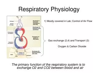
Gas Transport • Oxyhemoglobin: HbO2 • Deoxyhemoglobin: HHb • Carbaminohemoglobin: HbCO2 External Respiration: Oxygen and Carbon Dioxide concentration is measured as a unit of pressure called partial pressure (p) Blood coming into the lungs (pulmonary artery-capillary) is deoxygenated blood= PO2 is 40 mm Hg. PCO2 is 45 mm Hg
Air in the alveoli: PO2= 105 mm Hg PCO2= 40 mm Hg Oxygen and Carbon dioxide are highly fat soluble and can diffuse through membranes with ease. As gases pass from the blood pass an alveolus gases will diffuse from areas of higher concentration to lower. (diffusion gradients) PO2 in blood after passing alveolus is 105 mm Hg PCO2 in blood after passing alveolus is 40 mm Hg
Internal respiration: Exchange of gases in the tissues. CO2 is a byproduct of cellular metabolism. PCO2 in tissue space: 45 mm Hg PO2 in tissue spaces: 40 mm Hg O2 will diffuse into tissue spaces (105) and CO2 will diffuse into blood (45)
Gas Transport at the Tissues • Carbon dioxide transported to and from the lungs and tissues in three ways: • Dissolved in plasma 7% • Chemically bound to hemoglobin (carbaminohemoglobin) 23% • As Bicarbonate in plasma (Reaction between carbon dioxide and water, catalyzed by carbonic anhydrase) pH buffer system. 70% of CO2 is transported this way. • Chloride shift (Chloride anions diffuse into RBCs to counteract bicarbonate anions leaving RBCs) • Process results in diffusion of Oxygen from RBC to tissues and from Carbon dioxide from tissues to RBCs • This process is reversed in the Lungs
Oxygen Transport • Molecular oxygen carried in blood or bound to hemoglobin • HbO2- hemoglobin bound to oxygen • HHb + O2 -- HbO2 + H+ • Hb can bind 4 oxygens; after first binding, there is a higher affinity for other 3 • Hemoglobin is fully saturated when all 4 heme sites bound to oxygen
Clinical corner • Eupnea - quiet breathing • Tachypnea - rapid breathing • Costal breathing - shallow Diaphragmatic breathing - deep Atelectasis - collapse or incomplete expansion of lungs • Cheyne-Stokes respiration - irregular breathing (increase/decrease in depth and rapidity)
Laryngitis - inflammation of the vocal cords • Pleurisy - inflammation of the pleura Infant respiratory distress syndrome (IRDS) - insufficient surfactant produced, surface tension forces collapse of the alveoli • Hypoxia - inadequate amount of oxygen is delivered to body tissues anemic - to few RBCs, or RBCs with inadequate hemoglobin stagnant - blood circulation is impaired or blocked interference with gas exchange • Hypercapnia - apnea (breathing cessation) increase in carbon dioxide levels in cerebrospinal fluid, causing pH to decrease, exciting chemoreceptors to increase rate of breathing (compensating) • Hypocapnia- low levels of CO2 in plasma and CSFdue to depth and rate of breath increase (hyperventilation)
Chronic Obstructive Pulmonary Disease (COPD), common features: 1- Patients with history of smoking 2- Dyspnea - difficult or labored breathing 3- Coughing and frequent pulmonary infection 4- Will develop respiratory failure • COPDs: Obstructive emphysema - permanent enlargement of the alveoli, deterioration of alveolar walls • Chronic inflammation leads to lung fibrosis (lungs lose their elasticity) • Victims sometimes called "pink puffers" - breathing is labored, but doesn't become cyanotic because gas exchange remains adequate until late in the disease • Chronic bronchitis - inhaled irritants lead to chronic excessive mucus production by the mucosa of lower respiratory passageways and inflammation and fibrosis of that mucosa • Victims sometimes called "blue bloaters" - hypoxia and carbon dioxide retention occur