Download
1 / 43
460 likes | 1.87k Vues
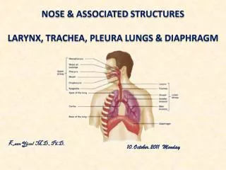
DEVELOPMENT OF LARYNX, TRACHEA AND BRONCHI. Dr. Mujahid Khan. Lower Respiratory Organs. The lower respiratory organs i.e. larynx, trachea, bronchi and lungs begin to form during the fourth week

E N D
DEVELOPMENT OF LARYNX, TRACHEA AND BRONCHI Dr. Mujahid Khan
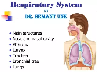
Lower Respiratory Organs • The lower respiratory organs i.e. larynx, trachea, bronchi and lungs begin to form during the fourth week • The respiratory primordiumالاولي ( في المرحله الجنينيه ) is indicated by a median وسط outgrowth from the caudal end النهايه السفليه of the ventral wall للجهه الاماميه of the primordial pharynx للبلعوم الاولي
Laryngotracheal Groove التجويف الداخلي • Is a primordium of the tracheobronchial tree • Develops caudal بشكل سفلي the fourth pair of pharyngeal pouches الحبيبات البلعوميه : هي التي تتطور لتعطي الراس والرقبه • The endoderm lining the laryngotracheal groove gives rise to bronchi and pulmonary epithelium
Respiratory Diverticulumتفرع جانبي يخرج من التفرع الاصلي • By the end of fourth week, the laryngotracheal groove has evaginatedتفرع جانبي to form a pouchجييب like respiratory diverticulum or lung bud تبرعم • Is located ventral بشكل خلفي to the caudal partللجزء السفلي of the foregut المعي الامامي • This diverticulum elongates and invested with splanchnicmesenchyme and its distal خلفي end enlarges to form a globular tracheal bud برعم
تنفصل عن بعضها بمرور الزمن حتى تكون المرئ والقصبه الهوائيه في النهايه
Tracheoesophageal folds & Septum • Longitudinal الطوليtracheoesophageal folds develop in the laryngotrachealdiverticulumكما في السهم في الشريحه السابقه • These folds approach each other and fuse to form a partition called tracheoesophageal septum • This septum divides the cranial علويpart of the foregut into a ventral امامي part, the laryngotracheal tube and a dorsal خلفي part, oropharynx and esophagus
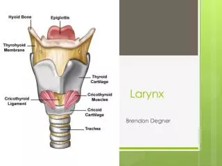
Development of Larynx • The epithelial lining of the larynx develops from endoderm of the cranial العلوي end of laryngotracheal tube • The cartilages of the larynx develop from the cartilages in the fourth and sixth pairs of pharyngeal arches العقد نفسها الحبيبات البلعوميه في الشريحه 3 • The laryngeal cartilages develop from the mesenchyme that is derived from neural crest cells
Development of Larynx • The laryngeal epithelium proliferates rapidly resulting in temporary occlusion سد مؤقت of the laryngeal lumen تجويف • Recanalizationاعاده فتح السدد of larynx normally occurs by the tenth week • :فائده هذا السدد وانفتاحه 1-Laryngeal ventricles بطين form during this recanalization • 2-These recesses تجويفات are bounded by folds of mucous membrane that become the vocal folds and vestibular folds
Epiglottis لسان المزمار • It develops from the caudal سفلي part of the hypopharyngealالجزء السفلي من البلعوم eminence بروز • The rostral part الجز الاوضح -الابرزof this eminence forms the posterior third or pharyngeal part of the tongue اللسان • Growth of the larynx and epiglottis is rapid during the first three years after birth • By this time the epiglottis has reached its adult form
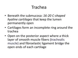
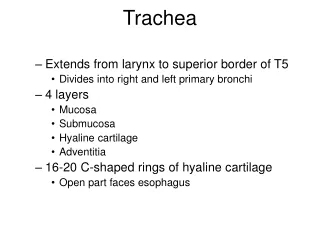
Development of Trachea • The endodermal lining of the laryngotracheal tube distal خلفي to the (larynx differentiates المتطوره )into the epithelium and glands of the trachea and pulmonary epithelium • The cartilages, connective tissue, and muscles of the trachea are derived from the splanchnicmesenchyme surrounding the laryngotracheal tube
Tracheoesophageal Fistula هو حاله تشوه جنيني لا يتم فيها تكوين المرئ و القصبه الهوائيه بشكل صحيح مما ينتج عنه وجود اتصال بينهما • It is an abnormal passage between the trachea and esophagus • Occurs once in 3000 to 4500 live births • Most affected infants are males مساكيييين • In more than 85% of cases, the fistula is associated with esophageal atresiaتضيق • It results from incomplete division of the cranial part of the foregut المعي into respiratory and esophageal parts
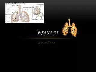
Development of Bronchi and Lungs • Tracheal bud بريعمات divides into two outpouchings, called primary bronchial buds انظر الصوره الصفحه القادمه • These buds grow laterally into the pericardioperitoneal canals, the primordiaالعضو الاولي of pleural cavities • Bronchial buds differentiate into the bronchi
Development of Bronchi and Lungs • Connection of each bronchial bud with the trachea enlarges to form the primordiumالعضو الاولي of a main bronchus • The right main bronchus is slightly larger than the left one and is oriented more vertically • The embryonic relationship persists دائم in the adult • The main bronchi subdivide into secondary bronchi that form lobar فصوص, segmental and intersegmental branches
اللون الزهري هو تكون الرئه ولاحظ عدد الفصوص يختلف من اليمنى لليسرى
Development of Bronchi and Lungs • The segmental bronchi, ten in right lung and eight or nine in the left lung begin to form by the seventh week • The surrounding mesenchyme also divides • Each segmental bronchus with its surrounding mass of mesenchyme is the primordiumالعضو الاولي of a bronchopulmonary segment جزء الرئه والشعيبات الموصله بها
Development of Bronchi and Lungs • By 24 weeks, about 17 orders of branches have formed and respiratory bronchioles have developed • An additional seven orders of airways develop after birth • As the bronchi develop, cartilaginous plates develop from the surrounding splanchnicmesenchyme
Development of Bronchi and Lungs • The bronchial muscle and pulmonary connective tissue and capillaries are also derived from this mesenchyme • As the lungs develop they acquire a layer of visceral pleura from splanchnicmesenchyme • The thoracic body wall becomes lined by a layer of parietal pleura derived from the somatic mesoderm
Maturation of the Lungs It is divided into four periods يتم على اربع مراحل: • Pseudoglandularالغدي الكاذب period • Canalicular period • Terminal saccular period • Alveolar period • تفصل بالشرائح القادمه
1- Pseudoglandular Period (6-16 weeks) • Developing lungs somewhat resembles تشبه an exocrine gland during this period • By 16 weeks all major elements of the lung have formed except those involved with gas exchange • Respiration is not possible • Fetuses born during this period cannot survive
2-Canalicular Period (16-26 weeks) • Cranial العلوي segments of the lungs mature faster than caudal السفلي ones • Lumina of bronchi and terminal bronchioles become larger • Lung tissue becomes highly vascular • By 24 weeks each terminal bronchiole has given rise to two or more respiratory bronchioles
Canalicular Period (16-26 weeks) • Each of the respiratory bronchioles divide into 3 to 6 tubular passages called alveolar ducts • Respiration is possible at the end of this period because some thin-walled terminal saccules (primordial alveoli) have developed at the end of respiratory bronchioles • Lung tissue is well vascularized • Fetus born at the end of this period may survive
3- Terminal Saccular period(26 weeks - birth) • Many more terminal saccules develop • Their epithelium becomes very thin • Capillaries begin to bulge into developing alveoli • By 26 weeks, the terminal saccules are lined by squamous epithelial cells of endodermal origin, type I pneumocytesخلايا مسؤوله عن تبادل الغازات
Terminal Saccular period(26 weeks - birth) • Gas exchange occurs across type I pneumocytes • Scattered تتناثر among the squamous epithelial cells are rounded secretory epithelial cells, type II alveolar cells or pneumocytesالمسؤؤوله عن الافراز • Type II pneumocytes secrete a mixture of phospholipids called surfactant راجع الفسيو • Surfactant forms as a monomolecular film over the internal walls of the terminal saccules
Terminal Saccular period(26 weeks - birth) • Surfactant production increases during the terminal stages of pregnancy • Surfactant reduces surface tension and facilitates expansion of the terminal saccules • Fetuses born prematurely at 24-26 weeks may survive if given intensive care • Fetuses may suffer from respiratory distress due to surfactant deficiency
Terminal Saccular period(26 weeks - birth) • Surfactant production begins by 20 weeks • By 26 – 28 weeks the fetus usually weighs about 1000 gm • Sufficient كافي terminal sacculesكيسسات هوائيه and surfactant ماده المانعه للاحتكاكare present to permit لتجعل من الممكنsurvival of a prematurely born infants • Pulmonary vasculature and sufficient surfactant are critical to the survival of the premature infant
4- Alveolar Period32 weeks – 8 years • Definition of the term alveolus depends on the change of terminal saccular period to alveolar period • Structures analogous to alveoli are present at 32 weeks saccules • The epithelial lining of the terminal sacs attenuates يضعف to an extremely thin squamous epithelial layer • Type I pneumocytes become so thin that the adjacent capillaries bulge into the terminal saccules
Alveolar Period32 weeks – 8 years • At the beginning of the alveolar period, each respiratory bronchiole terminates in a cluster عنقود of thin-walled terminal saccules, separated from one another by loose connective tissue • These terminal saccules represent future alveolar ducts
Alveolar Period32 weeks – 8 years The transition from dependence on the placenta for gas exchange to autonomous gas exchange requires the following adaptive changes in the lungs: • Production of adequate surfactant in the alveoli • Transformation of the lungs from secretory into gas exchanging organs • Establishment of parallel pulmonary and systemic circulations
Alveolar Period32 weeks – 8 years • Characteristic mature alveoli do not form until after birth • 95% of alveoli develop postnatallyبعد الولاده • Most increase in the size of the lungs results from an increase in the number of respiratory bronchioles and primordial alveoli rather than from an increase in the size of the alveoli
Alveolar Period32 weeks – 8 years • From third to eight year or so, the number of immature alveoli continues to increase • Unlike mature alveoli, immature alveoli have the potential for forming additional primordial alveoli • About 50 million alveoli, one sixth of the adult number are present in the lungs of a full-term newborn infant
Alveolar Period32 weeks – 8 years • By about the eighth year, the adult complement of 300 million alveoli is present • Breathing movements occur before birth, exerting جهد sufficient كافي force to cause aspiration شفط of some amniotic fluid into the lungs الجنين يتقيرد وياحول يتنفس ببطن امه فيدخل بعض من السائل المحيط به بالرئه بس هذا طبيعي (: • These breathing movements are not continuous and are detected by real-time ultrasonography
Alveolar Period32 weeks – 8 years • By birth the fetus has had the advantage of breathing exercise for months • These movements stimulate lung development • At birth the lungs are half filled with fluid derived from the amniotic cavity, lungs and tracheal glands
Alveolar Period32 weeks – 8 years The fluid in the lungs is cleared at birth by three routes: • Through the mouth and nose by pressure on the fetal thorax during delivery • Into the pulmonary capillaries • Into the lymphatics and pulmonary arteries and veins
Alveolar Period32 weeks – 8 years • Near term the pulmonary lymphatic vessels are relatively larger than in the adults • Three factors are important for normal lung development: • Adequate thoracic space for lung growth • Fetal breathing movements • Adequate amniotic fluid volume
Thank youقولوا لهذا الدكتور لا عمره يكتب محاضرات لحسه ...