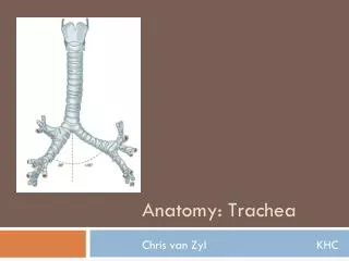
Trachea
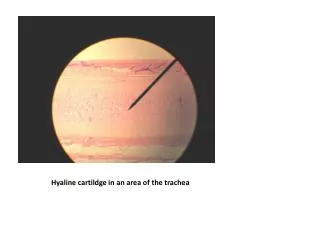
Trachea. Beneath the submucosa: 16-20 C-shaped hyaline cartilages that keep the lumen permanently open Cartilages form an incomplete ring around the trachea

Trachea
E N D
Presentation Transcript
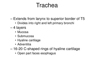
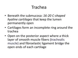
Trachea • Beneath the submucosa: 16-20 C-shaped hyaline cartilages that keep the lumen permanently open • Cartilages form an incomplete ring around the trachea • Open on the posterior aspect where a thick layer of smooth muscle fibers (trachealis muscle) and fibroelastic ligament bridge the open ends of each cartilage
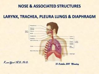
Lungs • Pair of conical organs that occupy the greater part of the thoracic cavity • Right lung – 3 lobes • Left lung – 2 lobes
Pleura • Double layer of fibrous tissue that envelopes the lungs • Parietal pleura • Visceral pleura • Pleural cavity • Mesothelium (simple squamous epithelium)
Bronchial Tree • The branching pattern of the main bronchi • Main bronchi • Right main bronchus • Left main bronchus • Secondary bronchi • Right – 3, Left – 2 • Tertiary bronchi • Right – 10, Left – 8
Bronchopulmonary Segment • Each tertiary bronchus and the area of the lung that it supplies • These segments each have their own artery (thus, each segment is supplied by a bronchus and an artery) • Each bronchopulmonary segment is a discrete anatomical and functional unit.
Bronchi • Extrapulmonary – main bronchi (before they enter lung substance) • Same structure as trachea, but smaller caliber • Intrapulmonary– all bronchi within the lung substance • Same structure as extrapulmonary, with exceptions
Bronchial Tree • Bronchioles – 1 mm diameter, disappearance of cartilage • Terminal bronchioles – 0.5 mm diameter or less • Respiratory bronchioles • Alveolar ducts
Bronchioles • Smaller caliber than bronchi • Wall: no cartilage, gland, lymph nodes • Epithelium: respiratory epithelium but diminishes in height and transforms to cuboidal as bronchial tree goes distally, no goblet cells • Smaller bronchioles: Clara cells
Clara Cells • Non-ciliated, columnar cells • Contain microvilli • Rounded apices • Contain dense secretory granules – protect the bronchiolar lining, form a non-sticky layer that helps keep the bronchioles patent