Download
1 / 34
350 likes | 504 Vues
Learn about the trachea and bronchi, their structure, location, blood supply, nerve supply, and relation to surrounding structures. Discover the branching patterns, histology, and function of the bronchial tree.

E N D
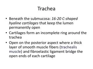
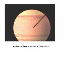
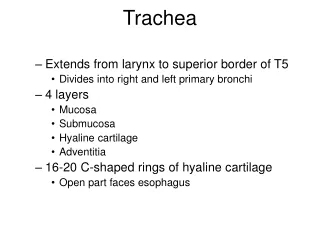
Trachea • Extends from larynx to superior border of T5 • Divides into right and left primary bronchi • 4 layers • Mucosa • Submucosa • Hyaline cartilage • Adventitia • 16-20 C-shaped rings of hyaline cartilage • Open part faces esophagus
Trachea • The trachea is a mobile cartilaginous and membranous tube • It begins in the neck as a continuation of the larynx at the lower border of the cricoid cartilage at the level of the sixth cervical vertebra • It descends in the midline of the neck
Trachea • In the thorax the trachea ends below at the carina by dividing into right and left principal (main) bronchi • During expiration the bifurcation rises by about one vertebral level • During deep inspiration may be lowered as far as the sixth thoracic vertebra
Trachea • In adults the trachea is about 4½ in. (11.25 cm) long and 1 in. (2.5 cm) in diameter • The fibroelastic tube is kept patent by the presence of U-shaped rings of hyaline cartilage embedded in its wall • The posterior free ends of the cartilage are connected by smooth muscle, the trachealis muscle
Relation • Anteriorly: The sternum, the thymus, the left brachiocephalic vein, the origins of the brachiocephalic and left common carotid arteries, and the arch of the aorta • Posteriorly: The esophagus and the left recurrent laryngeal nerve
Relation • Right side: The azygos vein, the right vagus nerve, and the pleura • Left side: The arch of the aorta, the left common carotid and left subclavian arteries, the left vagus and left phrenic nerves, and the pleura
Blood Supply • The upper two thirds are supplied by the inferior thyroid arteries • The lower third is supplied by the bronchial arteries
Lymph Drainage • The lymph drains into the pretracheal and paratracheal lymph nodes and the deep cervical nodes
Nerve Supply • The sensory nerve supply is from the vagi and the recurrent laryngeal nerves • Sympathetic nerves supply the trachealis muscle
Bronchi • Right and left primary bronchus goes to right lung • Carina – internal ridge • Most sensitive area for triggering cough reflex • Divide to form bronchial tree • Secondary lobar bronchi (one for each lobe), tertiary (segmental) bronchi, bronchioles, terminal bronchioles • Structural changes with branching • Mucous membrane changes • Incomplete rings become plates and then disappear • As cartilage decreases, smooth muscle increases • Sympathetic ANS – relaxation/ dilation • Parasympathetic ANS – contraction/ constriction
Bronchi • The trachea bifurcates behind the arch of the aorta into the right and left principal (primary, or main) bronchi • The bronchi divide into several million terminal bronchioles that terminate in one or more respiratory bronchioles
Wider, shorter, and more vertical than the left trachea Right Primary Bronchus Left primary bronchus Both primary bronchi have the same anatomic structure as the trachea.
Bronchi • Each respiratory bronchiole divides into 2 to 11 alveolar ducts that enter the alveolar sacs • The alveoli arise from the walls of the sacs as diverticula
The primary bronchi divide to form SECONDARY BRONCHI (lobar bronchi). • There is one secondary bronchus for each lobe of the lungs. • There are 2 lobes on the left lung. • There are 3 lobes on the right lung. • These also have the same anatomy as the trachea.
The secondary bronchi branch to form TERTIARY BRONCHI. • They continue to branch. • As they get smaller, they lose their cartilage. • When they lose their cartilage, they are called BRONCHIOLES which are microscopic.
The bronchioles terminate in the ALVEOLI through an ALVEOLAR DUCT. • The walls of the alveoli are one-cell thick and is covered in capillaries. • The alveoli are the functional unit of the lungs.
There are air sacs, where gas exchange occurs. • Walls of the alveoli are highly vascularized. • The alveoli are the terminal branches of the BRONCHIAL TREE. This arrangement allows for a drastic increase in surface area.
Bronchial tree • A highly branched system of air-conducting passages that originate from the left and right primary bronchi. • Progressively branch into narrower tubes as they diverge throughout the lungs before terminating in terminal bronchioles. • Incomplete rings of hyaline cartilage support the walls of the primary bronchi to ensure that they remain open. • Right primary bronchus is shorter, wider, and more vertically oriented than the left primary bronchus. • Foreign particles are more likely to lodge in the right primary bronchus.
Bronchial tree • The primary bronchi enter the hilus of each lung together with the pulmonary vessels, lymphatic vessels, and nerves. • Each primary bronchus branches into several secondary bronchi (or lobar bronchi). • The left lung has two secondary bronchi.The right lung has three secondary bronchi. • They further divide into tertiary bronchi. • Each tertiary bronchus is called a segmental bronchus because it supplies a part of the lung called a bronchopulmonary segment.
Bronchial Tree • Secondary bronchitertiary bronchibronchiolesterminal bronchioles • with successive branching amount of cartilage decreases and amount of smooth muscle increases, this allows for variation in airway diameter • during exertion and when sympathetic division active bronchodilation • mediators of allergic reactions like histamine bronchoconstriction • epithelium gradually changes from ciliated pseudostratified columnar epithelium to simple cuboidal epithelium in terminal bronchioles
Conduction vs. Respiratory zones • Most of the tubing in the lungs makes up conduction zone • Consists of nasal cavity to terminal bronchioles • The respiratory zone is where gas is exchanged • Consists of alveoli, alveolar sacs, alveolar ducts and respiratory bronchioles
Respiratory Bronchioles, Alveolar Ducts, and Alveoli • Lungs contain small saccular outpocketings called alveoli. • They have a thin wall specialized to promote diffusion of gases between the alveolus and the blood in the pulmonary capillaries. • Gas exchange can take place in the respiratory bronchioles and alveolar ducts as well as in the alveoli, each lung contains approximately 300 to 400 million alveoli. • The spongy nature of the lung is due to the packing of millions of alveoli together.
Bronchial “tree” and associated Pulmonary arteries 33
This “air-blood barrier” (the respiratory membrane) is where gas exchange occurs Oxygen diffuses from air in alveolus (singular of alveoli) to blood in capillary Carbon dioxide diffuses from the blood in the capillary into the air in the alveolus 34