TRACHEA
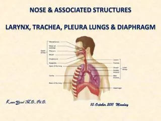
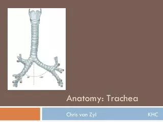
TRACHEA. What is Trachea. bony tube that connects the nose and mouth to the lungs. Anatomy. cartilaginous and membranous tube extends from the lower part of the larynx (6 th CV) to the upper border of the fifth thoracic vertebra divides into two bronchi

TRACHEA
E N D
Presentation Transcript
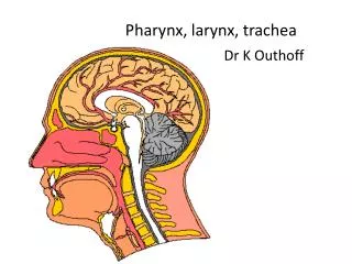
What is Trachea • bony tube that connects the nose and mouth to the lungs
Anatomy • cartilaginous and membranous tube • extends from the lower part of the larynx (6th CV) to the upper border of the fifth thoracic vertebra • divides into two bronchi • nearly but not quite cylindrical, being flattened posteriorly
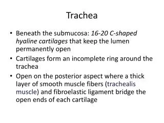
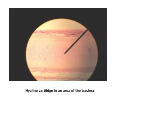
Anatomy • 11 cm. in length; 2 to 2.5 cm. in diameter • composed of 16 to 20 “c” shaped rings of cartilage connected by ligaments, with a ciliated-lined mucus membrane • always greater in the male than in the female • child vs adult • smaller • more deeply placed • and more movable than in the adult
Function • to maintain and protect the air way • mucus glands humidify the air as it passes through it and catches the small particles before they enter • trachea is supplied by nerves that are part of the cough reflex that helps get rid of irritation • coughing reflex allows the ciliated cells to push the object out of the trachea and out of the respiratory system.
Foreign Body • Epidemiology: bimodal - at the extremes of age • diagnosis is often missed initially • aspirated foreign bodies most commonly are lodged in the right main stem and lower lobe • history leads to diagnosis in most cases
Foreign Body • History • adults: respiratory distress associated with sedation from drugs, alcohol, or trauma; after medical procedures such as sedation or intubation; after facial trauma; with decreased ability to handle secretions • children: sudden paroxysms of coughing, sudden choking after eating or choking and/or coughing when a known, small object or food particle is within reach of the child
Foreign Body • Physical Examination • inspiratory stridor or expiratory wheezing • prolongation of the expiratory phase • medium-to-coarse rhonchi • tachypnea, nasal flaring, intercostal, subcostal, and suprasternal retractions • differences in percussion between hemithoraces *stridor indicates a partial upper airway or tracheal occlusion and is an ominous sign
Foreign Body • Management • initial supportive therapy: oxygen, cardiac monitor, pulse oximetry, and intravenous line • stridorous patients: racemic epinephrine via a nebulizer • extraction by bronchoscopy is the treatment of choice for tracheal foreign bodies • preoperative steroids and antibiotics may reduce complications such as airway edema and infection.
Tracheomalacia • supporting structure of the trachea is too floppy • weakness of the tracheal walls • expiratory stridor • during heavy breathing, the membranous posterior wall advances anteriorly, where it may approach or even touch the cartilaginous anterior tracheal wall, thus markedly reducing the airway lumen • symptoms are more prominent in an older infant as the respiratory movement increases • therefore, posterior tracheal wall migration increases as well • as a result, the affected infant may hyperextend his neck in order to adequately keep his airway open
Tracheomalacia • Primary tracheomalacia: congenital disorder • Secondary tracheomalacia: acquired disorder • cartilage weakness results from chronic wearing from some external pressure • Diagnosis: rigid bronchoscopy with the patient spontaneously ventilating • positive pressure ventilation from the respirator may balloon the trachea, thus hiding its floppiness • Treatment: supportive - aimed at preventing atelectasis • severe cases: tracheotomy, CPAP, or both
TracheoEsophageal Fistula • most common form (85%) is proximal esophageal atresia with a distal TEF • next most common type (4%) is the H type TEF without esophageal atresia • other types: proximal esophageal atresia with a proximal and distal TEF (2%), proximal esophageal atresia with a proximal TEF (1%) isolated esophageal atresia without a TEF (8%) • associated with trisomy 18, trisomy 21, and the VATER
TracheoEsophageal Fistula • symptoms: aspiration, dyspnea, and frank respiratory distress during feeding • diagnosis: failure to pass a nasogastric tube • a barium swallow study may reveal the fistula. • during rigid bronchoscopy, the fistula entrance may be observed in the posterior tracheal wall • cannulation of the fistula may facilitate surgical repair
Tracheal Stenosis • idiopathic or result from trauma • symptoms: dyspnea, stridor,initially diagnosed with asthma • treatment: • tracheal dilation using rigid bronchoscope • effect of dilation typically lasts from few days to 6 months • tracheal resection is so far the best alternative to cure the stenosis completely. • for stenosis of length greater than 5 cm a stent may be required to join the sections.