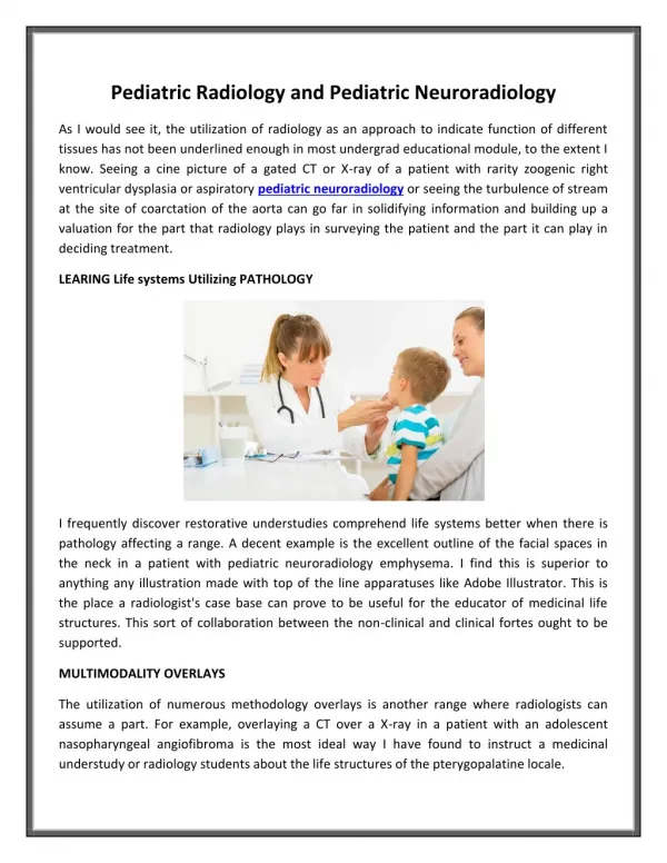
Pediatric CXR
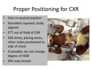
Pediatric CXR. Moritz Haager Nov 20, 2003. Not just small adults. What’s different about the pediatric CXR? Thymus Occult FB aspiration Congenital anomalies Smaller airways; more subtle disease findings Infectious etiologies & presentations. Normal newborn chest.

Pediatric CXR
E N D
Presentation Transcript
Pediatric CXR Moritz Haager Nov 20, 2003
Not just small adults • What’s different about the pediatric CXR? • Thymus • Occult FB aspiration • Congenital anomalies • Smaller airways; more subtle disease findings • Infectious etiologies & presentations
Thymus • Anterior upper mediastinal structure • Low density – should be able to see pulmonary vasculature behind it • Does not displace trachea posteriorly • Can increase in size after acute illness • Does not decrease in size w/ aging – rather stays roughly same size (~3x3 cm) and so becomes smaller in proportion to chest
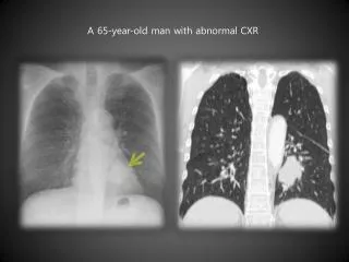
2-month old male with fever, noisy breathing, and tachypnea congestive heart failure due to congenital heart disease
CHF Findings • Cardiothoracic Ratio • Infants (less than 1 yo) • May be up to 0.6 • Toddlers and older • Maximum is 0.5 • CHF findings • Plethora, fluid in fissures, peribronchial cuffing • Don’t see pleural effusions and upper lobe redistribution as much as in adults
2 mo w/ VSD now presents with resp distress and “seizure” Cardiomegaly and absence of the thymic shadow most consistent with DiGeorge Syndrome (thymic and hypoparathyroid aplasia or hypoplasia).
11-month old female presents with fever and coughing. viral pneumonia
Viral Pneumonia • Far more common than bacterial pneumonia in kids • Non-specific X-ray findings: • Overexpansion • Peribronchial thickening • Interstitial infiltrates • Perihilar flaring
6-week old female with fever and cold symptoms. VS T39.1, P125, R45, BP 75/35, O2 sat 98% RA consolidation or atelectasis in the posterior segment of the right upper lobe
15-month old male with fever and coughing. right upper lobe consolidation.
15-month old male with fever, coughing, and tachypnea. Right middle and left lower lobe infiltrates
9 year old male with a history of fever, headache, nausea, and coughing round pneumonia
Round Pneumonia • Often present w/ pleuritic chest pain • See spherical opacity with poorly defined margins (compared to tumors which have clear margins) • Usually located posteriorly adjacent to pleura • Uncommon to see air bronchograms
10 year old male with a history of coughing and fever. Left-sided pneumonia and foreign body in the left mainstem bronchus
4-month old with respiratory distress and diminished breath sounds on the right Congenital RML lobar emphysema
Foreign Body Aspiration • Many FB’s are radiolucent • CXR less than perfect in detecting FB’s • Difficult histories often but if clear history of object in mouth + choking then need bronchoscopy regardless of radiographic results (especially with nuts) • If unclear and child asymptomatic then reasonable to d/c after informing parents of signs + Sx to be vigilant for
7 mo native child w/ cough & fever Miliary TB
11-month old female w/ near-drowning episode Pulmonary edema
13 month old male with wheezing, coughing, and rhinorrhea for the past month. T 37.5, P 138, RR 52, BP 95/40, O2 95% RA
Pulmonary Sequestration • Congenital non-functional (not connected to tracheobronchial tree) accessory lung tissue w/ anomalous arterial supply • Intralobar • More common, shares pleural lining w/ lung • Present w/ recurrent pneumonia • Extralobar • Contained in its own pleural sac • Present w/ resp distress or feeding difficulty in infancy • Angiography is gold standard for Dx • Multiple variants • Scimitar syndrome, horseshoe lung, cystic adenomatoid malformation, pulmonary AVM
cystic mass posterior to the trachea and the mainstem bronchi
Vascular Ring • Aortic arch malformation causing trachea & esophagus to be encircled by major blood vessels or branches thereof • See a right-sided aortic arch in nearly all cases • passes over the R mainstem bronchus rather than the L • Pushes carina to L rather than the usal R • May see only compression in double arch
5 year old, male w/ fever x 10 d, coughing, sore throat and mild back pain Lung abscess
Two week old male infant w/ a history of noisy breathing and worsening respiratory distress. VS T36.7, P160, R60, O2 sat 86% RA Congenital lobar emphysema.
References • Cases courtesy of: • Loren G. Yamamoto, MD, MPHAlson S. Inaba, MDRobert M. DiMauro, MDKapiolani Medical Center For Women And ChildrenDept. Pediatrics, University of Hawaii John A. Burns School of Medicine1319 Punahou Street, Honolulu, HI 96826http://www.hawaii.edu/medicine/pediatrics/pemxray/pemxray.html