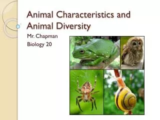
ANIMAL EVOLUTION AND DIVERSITY
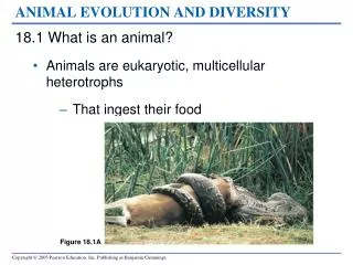
Figure 18.1A. ANIMAL EVOLUTION AND DIVERSITY. 18.1 What is an animal? Animals are eukaryotic, multicellular heterotrophs That ingest their food. 18.2 The ancestor of animals was probably a colonial, flagellated protist Cells in these protists Gradually became more specialized and layered.

ANIMAL EVOLUTION AND DIVERSITY
E N D
Presentation Transcript
Figure 18.1A ANIMAL EVOLUTION AND DIVERSITY • 18.1 What is an animal? • Animals are eukaryotic, multicellular heterotrophs • That ingest their food
18.2 The ancestor of animals was probably a colonial, flagellated protist • Cells in these protists • Gradually became more specialized and layered Somaticcells Digestive cavity Reproductivecells 2Hollow sphere of unspecialized cells (shown in cross section) 3Beginning of cell specialization (cross section) 4Infolding (cross section) 1Colonial protist, an aggregate of identical cells 5Gastrula-like “proto-animal” (cross section) Figure 18.2A
Top Dorsal surface Anterior end Posterior end Ventral surface Bottom Figure 18.3A • 18.3 Animals can be characterized by basic features of their “body plan” • Animal body plans • May vary in symmetry
Tissue-filled region (from mesoderm) Body covering (from ectoderm) Digestive tract (from endoderm) Body covering (from ectoderm) Muscle layer (from mesoderm) Digestive tract (from endoderm) Pseudocoelom Body covering (from ectoderm) Coelom Tissue layer lining coelomand suspendinginternal organs(from mesoderm) Digestive tract(from endoderm) • Vary in body cavity Figure 18.3B–D
Molluscs Annelids Sponges Flatworms Chordates Arthropods Cnidarians Nematodes Echinoderms Deuterostomes Protostomes Bilaterians Radial symmetry Bilateral symmetry Eumetazoans No true tissues True tissues Figure 18.4 Ancestral colonial protist • 18.4 The body plans of animals can be used to build phylogenetic trees • One hypothesis of animal phylogeny • Is based on morphological comparisons
Figure 18.5A–C INVERTEBRATES • 18.5 Sponges have a relatively simple, porous body • Sponges, phylum Porifera • Are the simplest animals and have no true tissues
Pores Choanocyte Amoebocyte Waterflow Skeletalfiber Centralcavity Flagella Choanocytein contactwith anamoebocyte Figure 18.5D • Flagellated choanocytes • Filter food from the water passing through the porous body
18.6 Cnidarians are radial animals with tentacles and stinging cells • Cnidarians, phylum Cnidaria • Have true tissues and radial symmetry
Figure 18.6A–C • 18.6 Cnidarians are radial animals with tentacles and stinging cells • phylum Cnidaria • Have true tissues and radial symmetry • Their two body forms are • Polyps, such as hydra • Medusae, the jellies • They have a gastrovascular cavity • And cnidocytes on tentacles that sting prey
Gastrovascularcavity Nerve cords Mouth Eyespots Nervoustissueclusters Figure 18.7A Bilateral symmetry • 18.7 Flatworms are the simplest bilateral animals: phylum Platyhelminthes • Are bilateral animals with no body cavity, and a simple nervous system
Units withreproductivestructures Scolex(anteriorend) HooksSucker Colorized SEM 80 Flukes and tapeworms Are parasitic flatworms with complex life cycles Figure 18.7B
Muscle tissue Trichinella juvenile Mouth Colorized SEM 400 LM 350 Figure18.8A, B • 18.8 Nematodes have a pseudocoelom and a complete digestive tract • Nematodes, phylum Nematoda • Have a pseudocoelom and a complete digestive tract • Are covered by a protective cuticle & free living
Visceral mass Reproductive organs Coelom Heart Kidney Digestive tract Mantle Shell Digestive tract Mantle cavity Radula Radula Anus Mouth Gill Mouth Foot Nerve cords Figure 18.9A • 18.9 Diverse molluscs are variations on a common body plan • All molluscs have a muscular foot and a mantle • Which may secrete a shell that encloses the visceral mass • Many mollusks • Feed with a rasping radula
Figure 18.9B, C • Gastropods • Gastropods are the largest group of molluscs • And include the snails and slugs
Figure 18.9D • Bivalves • The bivalves have shells divided into two halves • And include clams, oysters, mussels, and scallops
Figure 18.9E, F • Cephalopods • Cephalopods are adapted to be agile predators • And include squids and octopuses
18.10 Annelids are segmented worms • The segmented bodies of phylum Annelida • Give them added mobility for swimming and burrowing
Epidermis Anus Circular muscle Segment wall (partition between segments) Segment wall Longitudinal muscle Dorsal vessel Excretory organ Mucus-secreting organ Intestine Bristles Bristles Dorsal vessel Coelom Nerve cord Ventral vessel Excretory organ Digestive tract Brain Segment wall Blood vessels Giant Australian earthworm Mouth Nerve cord Pumping segmental vessels • Earthworms and Their Relatives • Earthworms • Eat their way through soil • Have a closed circulatory system Figure 18.10A
Figurer 18.10D Figure 18.10B, C • Annelids: Segmented Worms • PolychaetesForm the largest group of annelids Search for prey on the seafloor or live in tubes and filter food particles
Cephalothorax Abdomen Thorax Antennae (sensory reception) Head Swimming appendages Walking legs Figure 18.11A Mouthparts (feeding) Pincer (defense) • 18.11 Arthropods are segmented animals with jointed appendages and an exoskeleton • The diversity and success of arthropods • Are largely related to their segmentation, exoskeleton, and jointed appendages
Colorized SEM 900 A black widow spider (about 1 cm wide) A dust mite (about 420 µm long) A scorpion (about 8 cm long) Figure 18.11B, C • Chelicerates • Chelicerates include • Horseshoe crabs • Arachnids, such as spiders, scorpions, mites, and ticks
Figure 18.11D • Millipedes and Centipedes • Millipedes and centipedes • Are identified by the number of jointed legs per body segment
Figure 18.11E • Crustaceans • The crustaceans • Are nearly all aquatic • Include crabs, shrimps, and barnacles
18.12 Insects are the most diverse group of organisms • Insects have a three-part body consisting of • Head, thorax, and abdomen • Three sets of legs • Wings (most, but not all insects)
Many insects undergo • Incomplete or complete metamorphosis
Abdomen Head Thorax Antenna Forewing Eye Mouthparts Hindwing • A. Order Orthoptera • The order orthoptera includes • Grasshoppers, crickets, katydids, and locusts Figure 18.12A
Figure 18.12B • B. Order Odonata • The order odonata includes • Dragonflies and damselflies
Figure 18.12C • C. Order Hemiptera • The order hemiptera includes • Bedbugs, plant bugs, stinkbugs, and water striders
Figure 18.12D • D. Order Coleoptera • The order coleoptera includes • Beetles
Figure 18.12E • E. Order Lepidoptera • The order lepidoptera includes • Moths and butterflies
Haltere Figure 18.12F • F. Order Diptera • The order Diptera includes • Flies, fruit flies, houseflies, gnats, and mosquitoes
Figure 18.12G • G. Order Hymenoptera • The order hymenoptera includes • Ants, bees, and wasps
Tube foot • 18.13 Echinoderms have spiny skin, an endoskeleton, and a water vascular system for movement • Echinoderms, phylum Echinodermata • Includes organisms such as sea stars and sea urchins • Are radially symmetrical as adults Tube foot Spine Figure 18.13B, C
Anus Spines Stomach Tube feet Canals Figure 18.13A • The water vascular system • Has suction cup–like tube feet used for respiration and locomotion
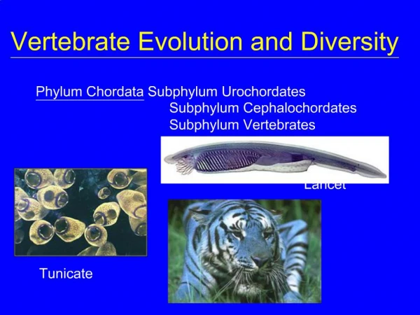
18.14 Our own phylum, Chordata, is distinguished by four features • Chordates, phylum Chordata have • A dorsal hollow nerve cord • A stiff notochord • Pharyngeal slits • A muscular post-anal tail
Excurrent siphon Post-anal tail Dorsal, hollow nerve cord Head Pharyngeal slits Notochord Mouth Mouth Muscle segments Pharynx Dorsal, hollow nerve cord Pharyngeal slits Notochord Digestive tract Water exit Post-anal tail Adult (about 3 cm high) Larva Segmental muscles Anus Figure 18.14A, B • The simplest chordates are tunicates and lancelets • Marine invertebrates that use their pharyngeal slits for suspension feeding
Chordates Craniates Vertebrates Jawed vertebrates Tetrapods Amniotes Lobe-fins Reptiles Lancelets Mammals Hagfishes Tunicates Lampreys Amphibians Milk Sharks, rays Ray-finned fishes Amniotic egg Legs Lobed fins Lungs or lung derivatives Jaws Vertebral column Head Brain Ancestral chordate VERTEBRATES • 18.15 Derived characters define the major clades of chordates • A chordate phylogenetic tree is based on a sequence of derived characters Figure 18.15
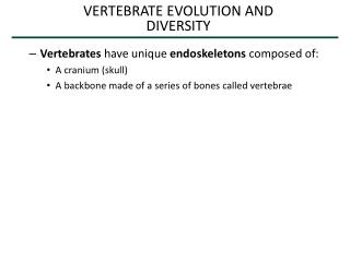
Most chordates are vertebrates • With a head and a backbone made of vertebrae
Figure 18.16A • 18.16 Lampreys are vertebrates that lack hinged jaws • Lampreys lack hinged jaws and paired fins
Skeletal rods Skull Gill slits Mouth Figure 18.16B • Most vertebrates have hinged jaws • Which may have evolved from skeletal supports of the gill slits
18.17 Jawed vertebrates with gills and paired fins include sharks, ray-finned fishes, and lobe-fins • Three lineages of jawed vertebrates with gills and paired fins • Are commonly called fishes
Figure 18.17A • Chondrichthyans • Chondrichthyans • Have a flexible skeleton made of cartilage • Include sharks and rays
Bony skeleton Dorsal fin Gills Anal fin Operculum Swim bladder Pectoral fin Pelvic fin Heart Rainbow trout, a ray-fin Figure 18.17B • Ray-finned Fishes • The ray-finned fishes have • A skeleton reinforced with a hard matrix of calcium phosphate • Operculi that move water over the gills • A buoyant swim bladder
Figure 18.17C • Lobe-fins • The lobe-fin fishes • Have muscular fins supported by bones
Bones supporting gills Tetrapod limb skeleton Figure 18.18A • 18.18 Amphibians were the first tetrapods—vertebrates with two pairs of limbs • Amphibians • Were the first tetrapods with limbs allowing movement on land
Figure 18.18B–D • Include frogs, toads, salamanders, and caecilians
Most amphibian embryos and larvae • Still must develop in water
Figure 18.19A, B • 18.19 Reptiles are amniotes—tetrapods with a terrestrially adapted egg • Terrestrial adaptations of reptiles include • Waterproof scales • A shelled, amniotic egg
Living reptiles other than birds • Are ectothermic