

SlideServe
Menu
Préférences
© SlideServe · v2026

ECG 1

clark-bartlett avatar
12 diapositives · 413 Views · Nov 14, 2014

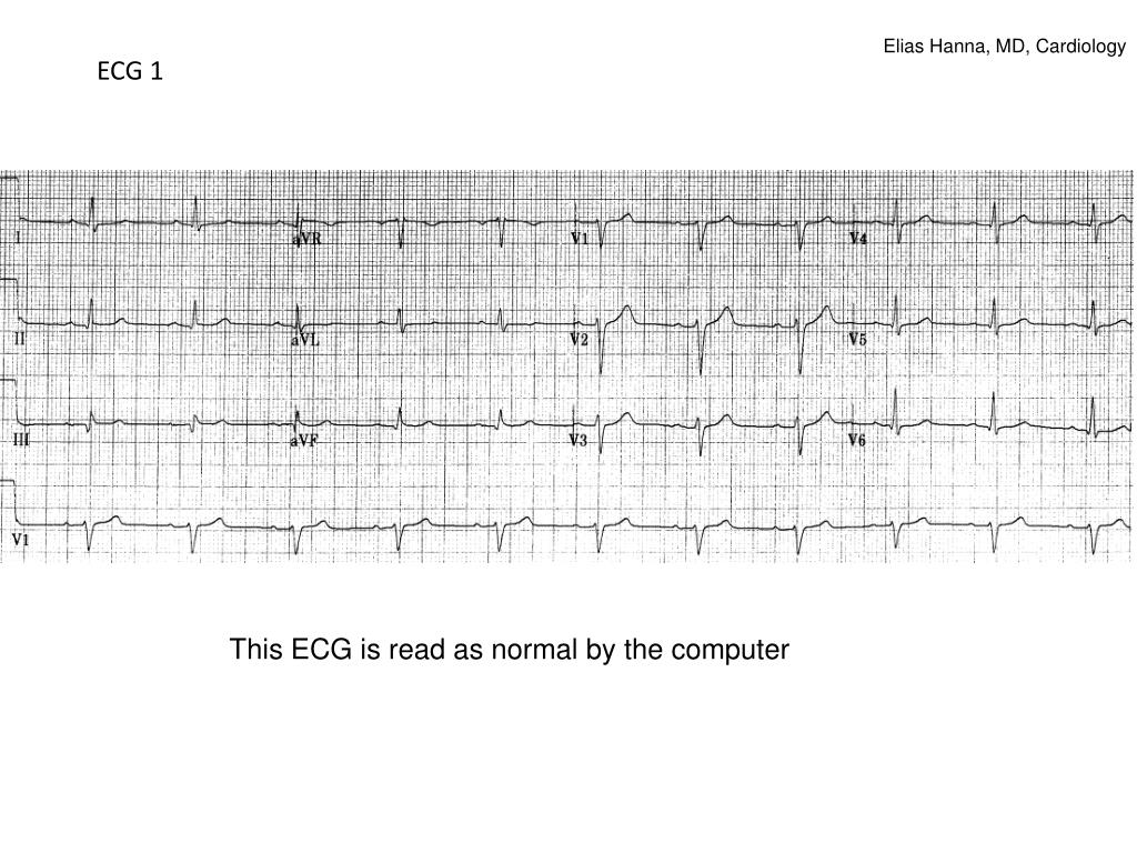
Elias Hanna, MD, Cardiology. ECG 1. This ECG is read as normal by the computer. Q wave that is part of a QR or QRS complex is abnormal if: *wide > 1 small box *or if present in V1-V3 to the right of the transition zone, regardless of how small it is.
ECG 1 - slide 1
01 / 12ECG 1
1 / 17

Télécharger la présentation

ECG 1
Une image / un lien ci-dessous est fourni (tel quel) pour télécharger la présentation

Politique de téléchargement : le contenu du site vous est fourni TEL QUEL pour votre information et votre usage personnel ; il ne peut être vendu, concédé sous licence ni partagé sur d’autres sites sans l’accord de son auteur.Le contenu vous est fourni TEL QUEL, pour votre information et votre usage personnel uniquement. Téléchargez la présentation en cliquant sur ce lien. Lors du téléchargement, si vous ne parvenez pas à télécharger une présentation, l’éditeur a peut-être supprimé le fichier de son serveur.Pendant le téléchargement, si vous ne parvenez pas à obtenir une présentation, le fichier a peut-être été supprimé par l’éditeur.

F I N

Plus de contenu associé

Transcription de la présentation
  1. Elias Hanna, MD, Cardiology ECG 1 This ECG is read as normal by the computer

  2. Q wave that is part of a QR or QRS complex is abnormal if: *wide > 1 small box *or if present in V1-V3 to the right of the transition zone, regardless of how small it is. Exception: Wide Q may be normally seen in leads III or aVL in an isolated fashion QS pattern is abnormal in all leads except III, and leads V1 +/- V2

  3. ECG 1 On this ECG, there is an abnormal Q in II, III, aVF diagnostic and specific for an inferior infarct of indeterminate age Although Q is not deep, it is wide > 1 box, signifying a pathological Q. Had the wide Q been isolated in lead III, this ECG would be considered normal Echo confirmed the old inferior infarct

  4. ECG 2 56 yom, presents 14 hrs after CP onset.

  5. ECG 2

  6. ECG 2 shows minimal residual ST elevation with a narrow tiny q in leads V3-V4 and post-ischemic T-wave abnormality (not true Wellens since there are Qs). Q wave of any size, when seen in the precordial leads before the transition zone as part of a qrS complex (i.e.,qrS before the transition zone), is abnormal Q and is 100% indicative of MI. This is basically late STEMI presentation at a time where ST elevation is resolving and Q waves/T inversion appeared.

  7. Q wave that is part of a QR or QRS complex is abnormal if: *wide > 1 small box *or if present in V1-V3 to the right of the transition zone, regardless of how small it is. Exception: Wide Q may be seen in leads III or aVL in an isolated fashion QS pattern is abnormal in all leads except III, and leads V1 +/- V2

  8. ECG 3 Inferior Q and anterolateral QS pattern (wide monophasic Q, arrows). QS in leads V1-V2 may be normal, but not in V3-V6

  9. Q wave that is part of a QR or QRS complex is abnormal if: *wide > 1 small box *or if present in V1-V3 to the right of the transition zone, regardless of how small it is. Exception: Wide Q may be seen in leads III or aVL in an isolated fashion QS pattern is abnormal in all leads except III, and leads V1 +/- V2

  10. On this ECG, QS is seen in V1-V2, small R wave is seen in V3-V6: as opposed to QS extending to V3 or beyond, QS limited to V1 or V2 is not definitely an anterior MI. Only 20% of pts with QS in V1-V2 without other abnormalities on the ECG have anterior MI.

  11. ECG 4 Inferior and posterior MI. Wide (> 1 small box) +/- tall R in V1 or V2 is indicative of posterior Q-wave MI

  12. Summary: Abnormal Q= *Q wider > 1 box (0.04 sec) and deeper > 1 box in 2 contiguous leads *Q of any width before the precordial transition zone (i.e., V1-V3) *Q may have a QS shape, which is a very wide and deep Q wave usually indicative of MI, except in leads III or V1 +/-V2 where it may be represent a normal variant *Wide +/- tall R in V1 or V2 indicates posterior MI