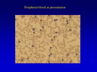
Peripheral blood
Peripheral blood. Practical lesson WS 10 – group 1051. Teacher: Tomáš Kučera. Preparation, staining and evaluation of the blood smear Blood cells are studied in smears prepared by spreading of a drop of peripheral blood in a thin layer on the microscopic slide. Procedure :


Peripheral blood
E N D
Presentation Transcript
Peripheral blood Practical lesson WS 10 – group 1051 Teacher: Tomáš Kučera
Preparation, staining and evaluation of the blood smear Blood cells are studied in smears prepared by spreading of a drop of peripheral blood in a thin layer on the microscopic slide. Procedure: Disinfection of the skin of the fingertip of the left fourth or third finger (at right-handers) Puncture into the ball of fingertip with sterile lancet or single-use needle The first drop of blood is wiped off because blood is diluted by tissue fluid The second dropof blood is placed near an end of a glass (microscopic) slide and spread using another slide (called “spreader” slide)
Spreading: spreader slide is moved over the glass slide at an angle of 45º, when slide edge touches the blood drop, the blood spreads by capillary forces along its edge. A thin film of blood is obtained by a smooth quick motion of the spreader slide across a glass slide. The air dried blood smear is fixed and stained. Blood smears are stained with mixtures of acidic (eosin) and basic dyes (methylene blue and its oxidation products – methylene violet and azure)
PAPPENHEIM´S METHOD Method is used for staining of peripheral blood and bone marrow smears Fixation of blood smear with May-Grünwald solution (eosinate of methylene blue in methanol) .......... 3 min. Stainingwith diluted May-Grünwald solution (the same amount of distilled water was added) ......... 1 – 2 min. Pour off mixture Stainingwith Giemsa-Romanowsky (eosinate of the methylene azure, blue and violet) …………….. 15 min. Washing (distilled water), air-drying Results of staining: red blood cells – pink/red,nuclei – purple/blue, neutrophilic granules – salmon pink, eosinophilic granules – brick-red, basophilic granules – blue/violet, cytoplasm oflymphocyte – sky blue, cytoplasm of monocyte – pale blue (grayish or greenish), azurophilicgranules - purple red
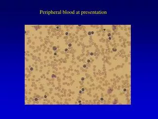
Evaluation of the blood smear Blood smear is observed in light microscope using HI objective (oil.im.,100x) andimmersion oil RBC evaluation: size, shape, structure anisocytosis (microcytosis, macrocytosis), poikilocytosis- variation in RBC shape: spherocytosis,ovalocytosis, sickle cells WBC evaluation: size and morphology Leukogram (differential leukocyte count) - proportional incidence (%) Arneth formula - reflects ratio of the neutrophilic granulocytes based on the number of nuclear lobes.Shift to left - predominance of the young forms (band and two lobes). Shift to right - predominance of the old forms (four and five lobes).
FORMED ELEMENTS - blood cell count (number per μL or L) ERYTHROCYTES - Male: 4.5-6.0x 10¹² / L Female:4.0-5.2x 10¹²/ L LEUKOCYTES – 4.000 -10.000 per μL (4 – 10 x 109 / L) LEUKOGRAM (differential leukocyte count) Neutrophils: 60-70 % (band form: 2-5 %) Eosinophils: 2-5 % Basophils: 0-1 % Lymphocytes: 20-40 % Monocytes: 2-10 % THROMBOCYTES - 150.000 -400.000 per μL
NG Ly
NG band form
Eos Ly
NG BG Mo