1 / 57

How to look at A peripheral blood smear

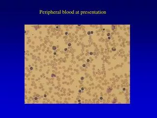
How to look at A peripheral blood smear. Low Power. 1- Scan the slide under low power (10x) 2- Find a good area 3- Evaluate red cell distribution 4- Evaluate the white count 1 WBC = 1000-2000mm 3. Too thick area. Red cells appear as; microcyte hypochrome rouleaux. Too thick area.

tisbell
Télécharger la présentation

How to look at A peripheral blood smear

An Image/Link below is provided (as is) to download presentation Download Policy: Content on the Website is provided to you AS IS for your information and personal use and may not be sold / licensed / shared on other websites without getting consent from its author. Content is provided to you AS IS for your information and personal use only. Download presentation by click this link. While downloading, if for some reason you are not able to download a presentation, the publisher may have deleted the file from their server. During download, if you can't get a presentation, the file might be deleted by the publisher.

E N D

Presentation Transcript


  1. How to look at A peripheral blood smear

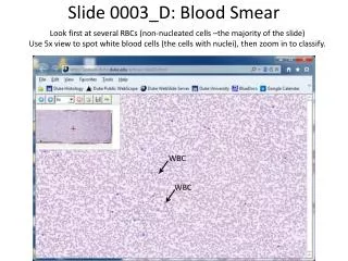
  2. Low Power 1- Scan the slide under low power (10x) 2- Find a good area 3- Evaluate red cell distribution 4- Evaluate the white count 1 WBC = 1000-2000mm3

  3. Too thick area Red cells appear as; • microcyte • hypochrome • rouleaux

  4. Too thick area

  5. Too thin Red cells appear as • macrocyte • spherocyte • poikilocyte

  6. Too thin area

  7. Good area

  8. What are you looking for at a peripheral smear (Oil immersion) • Red cell morphology • Platelet number and morphology • WBC differential and morphology • Blood parasite

  9. RBC MORPHOLOGY

  10. Size Shape Color Inclusion Distribution

  11. Evaluate red cells for size

  12. Normocytes

  13. Microcytes

  14. Macrocytes

  15. Macro-ovalocytes

  16. Anisocytosis

  17. color Hb content & distribution

  18. Normochrome

  19. Hypochrome

  20. spherocyte

  21. Microspherocyte

  22. Dimorphic population

  23. Target cells

  24. Stomatocytes

  25. shape

  26. Normocyte

  27. Tear-drop cells

  28. Acanthocytes

  29. Echinocytes

  30. Ovalocyte - Elliptocyte

  31. Sickle cells

  32. Keratocyte (Horn cell)

  33. Pincher cells

  34. Schistocytes

  35. Distribution

  36. Erythrocyte antibody rosetting (EA-rosetting)

  37. Rouleaux formation

  38. Inclusions

  39. HbH(Golf ball)

  40. HbC crystal

  41. Basophilic stippling

  42. Howell Jolly bodies

  43. Pappenheimer bodies

  44. Cabot ring

  45. babesia

  46. Nucleated red blood cell (NRBC)

More Related