Download
1 / 19
240 likes | 875 Vues
Staining of Blood Smear. Romanowsky stain. Romanowsky stain → Eosin Y and Azure B) Eosin:Acidic Dye bind to Basic groups (Hb,Granules) → reddish or orange color Azure B: Dye bind to nucleic acid & nucleoproteins → Blue-violet color

E N D
Romanowsky stain • Romanowsky stain→Eosin Y and Azure B) Eosin:Acidic Dye bind to Basic groups (Hb,Granules) → reddish or orange color Azure B: Dye bind to nucleic acid & nucleoproteins →Blue-violet color Fixation →Methanol<4%water, with 1 hour Delay : Adherence of Pro. To slide → Blue background
Romanowsky stain • Wright • Wright – Giemsa • Lishman • May- grunwald - Giemsa • jenner
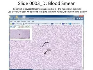
Making blood film • Blood film can be prepared from fresh blood without anticoagulant or from EDTA anticoagulanted blood. • blood film should be made on clean glass . • Clear without any dust
Wedge method • The most commonly in routine lab • Method • Thickness or thinness regulated by • Amount of blood • Speed of spreader • Angle
Optimal blood smear characteristic • minimum 2.5 cm in length terminating at least 1 cm from the end of the slide • Gradual Transition in thickness from thick to thin area ending in a Square or straight edge • No streaks , waves , or troughs
Spinner • Blood films that combine the advantages of easy handling of the wedge slide & uniform distribution of cells of the coverglass reparation • Method • Advantages: minimal exposure to biohazardous , increased optimal counting area
Reference method • Pure Azure B (260mg/100ml methanol) • Pure eosin y ( 130 mg/100ml methanol) • 1 part Azure B + 1 part eosin y +10 part sorensens phosphate buffer 66mmol/l ph= 6.8 • 10 min • washing
Quality Control رنگ پس از تهيه از نظر آلودگی قارچی و ميکروبی وهر گونه رسوب و پارتيکل وهمچنين نحوه رنگ گرفتن سلول های خونی بررسی می گردد.رنگ آميزی گسترش های خونی روتين نيز هفته ای يک بار توسط مسئول فنی داخلی از نظر موارد فوق بررسی می گردد که بصورت مکتوب ومستند بايد در آزمایشگاه قرار گيرد.کيفيت رنگ آميزی مورد قبول سلول ها مطابق جدول زير می باشد .