Download

1 / 14
140 likes | 317 Vues
Packed Cell Volumes Total Proteins Blood Smear Prep. PCV (Packed Cell Volume). In the CBC, we determine the number of RBC’s in several different ways. The quickest and easiest is called the hematocrit, also referred to as the packed cell volume (PCV).

E N D
PCV (Packed Cell Volume) • In the CBC, we determine the number of RBC’s in several different ways. The quickest and easiest is called the hematocrit, also referred to as the packed cell volume (PCV). • The hematocrit or the packed cell volume will tell you if the animal is dehydrated or anemic.
Whole blood is collected in an anticoagulant, EDTA, and placed in a capillary tube. Microhematocrit tubes should be filled to the designated line, with one ended plugged with clay sealant
A blood sample should be spun in a microhematocrit centrifuge for 2-5 minutes. • Lie the tube in the centrifuge with plugged end to the outside. *Note the number of your slot. Ensure that a balancing hematocrit tube is placed opposite, either by someone placing their tube there, or by adding an empty tube • The cells are heavier than the plasma and are compacted at one end of the tube.
The bottom of the RBC layer should be at the zero line and the top of the plasma on the top line (page 37 Fig 2-7) • PCV is determined as the percentage of the cellular portion relative to the total amount of blood in the tube
Plasma Evaluation • Plasma color and transparency may be helpful in the determination of a diagnosis and should be recorded . • Normal plasma is clear and a pale straw –yellow color • Cloudy Serum = lipemic • Reddish Tinge = hemolyzed • Yellow = Icteric (liver dz) • If the serum is anything but clear, a falsely elevated total protein will result. • Page 36
Plasma Protein Concentration or Total Protein / Total Solids • Plasma Protein concentrations estimation by refractometryis an important component of the CBC in all species • The plasma used to determinate the TP is collected by breaking the hematocrit tube just above the buffy coat- plasma interface.
The plasma is allowed to flow onto the refractometer prism. • Page 37 fig 2-9
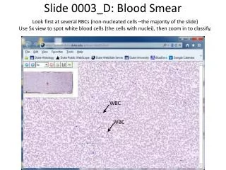
Blood Film / Smears • The blood film is used to perform the differential WBC count; estimate platelet numbers; and evaluate the morphological features of WBCs, RBCs and platelets. • Wedge smears are prepared by placing a small drop of blood on a clean glass microscope slide
Always stain using the lightest to darkest stain. • Dip each slide 10-12 times • Remember which side of your slide is up. • Rinse off stain using a gentle stream of water. • Allow slide to air dry.