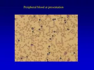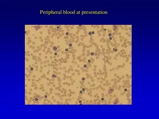
Peripheral Blood
Blood-Hematopoiesis-Lymphatics. Peripheral Blood. William F. Kern, M.D. Director, Laboratory Hematology Department of Pathology william-kern@ouhsc.edu.

Peripheral Blood
E N D
Presentation Transcript
Blood-Hematopoiesis-Lymphatics Peripheral Blood William F. Kern, M.D. Director, Laboratory Hematology Department of Pathologywilliam-kern@ouhsc.edu
Downloading or copying any of the photographs, images or diagrams from this presentation for any purpose other than studying for BHL is prohibited
Blood: Constituents • Plasma (liquid) • Cells: • Erythrocytes (Red Blood Cells) • Leukocytes (White Blood Cells) • Platelets
Anticoagulated Blood Sample Allowed to Settle Plasma (50-60%) T Buffy Coat (WBCs & Platelets) R Erythrocytes (40-50%) R T Packed Cell Volume (“Hematocrit”) = x 100 [%]
Plasma • Plasma = Liquid phase of anticoagulated blood • ~90% Water • Proteins: • Albumin • Coagulation factors • Immunoglobulins, lipoproteins, others • Ions, glucose, amino acids, waste products Note: Serum = fluid remaining after blood clotted
ErythrocytesRed Blood Cells (RBCs) • Primary function: Oxygen transport • Hemoglobin = Oxygen carrying molecule • Oxygen bound to iron in heme ring • Secondary function: Acid-base balance • Anucleate • Life span ≈ 120 days • Most phagocytized & destroyed in spleen
Hemoglobin • Heme = iron atom in protoporphyrin ring • Globin = protein portion • Hemoglobin = tetramer: 2 a-chains, 2 b-type chains: • Hb A: a2b2>95%* • Hb A2: a2d2 1.7-3.3%* • Hb F: a2g2 <2%* * Normal adult values at OUMC
ErythrocytesRed Blood Cells (RBCs) • Reddish-orange color on blood smear • Biconcave disk shape • Central pallor: ~ 1/3rd diameter of RBC • Diameter: ~ 7-8 microns • ~ Same size as nucleus of small, resting lymphocyte
Erythrocytes Erythrocyte Erythrocyte Lymphocyte
Erythrocytes: Descriptive Terms • Normocytic: Normal size • Microcytic: Small (<6.5 microns) • Macrocytic: Large (>8.5 microns) • Normochromic: Normal central pallor • Hypochromic: Widened central pallor (>1/2 diameter of cell)
Reticulocytes • Reticulocyte = immature RBC containing RNA • Contain RNA for ~1 day after leaving marrow • Normal reticulocyte count ~ 1% of RBCs (~1% of RBCs replaced daily) • Basophilic (bluish) on routine blood smear stains • Definitive identification requires special stain (“reticulocyte stain”)
Reticulocytes Reticulocyte
Erythrocytes: Normal Parameters (Note: Values vary between different laboratories) MCV = Mean Corpuscular Volume (Femtoliters [10-15 L])
Leukocytes:White Blood Cells (WBCs) • Granulocytes: Contain specific granules • Neutrophils (polymorphonuclear leukocytes or “Segs”) • Eosinophils (red granules) • Basophils (dark blue granules) • Agranulocytes: May contain lysosomes (nonspecific granules): • Lymphocytes • Monocytes
Neutrophils (“Polys”) • Most common type of leukocytes • Function: Phagocytosis, predominantly bacteria • Appearance: • Nucleus segmented (“Seg”) or horseshoe-shaped (“Band”) • Granules small, weakly stained (“neutrophilic”) Neutrophils are your main defense against bacterial infection
Segmented Neutrophil(PMN; “Seg” or “Poly”) • Nucleus with distinct lobes connected by thin strands of chromatin • Finely granular cytoplasm • Weakly staining granules
Band Neutrophil (“Band”) • Non-segmented, horseshoe-shaped nucleus • Less mature than segmented neutrophil • Fully functional • Count towards absolute neutrophil count
Neutrophils (“Polys”) • Lifespan: ~10 hours in circulation, 1-4 days in tissue • Increase in acute infection, inflammation or other stress: • Increase in “band” forms particularly suggests acute infection or inflammation
Eosinophils (“Eos”) • Large, intensely red (“eosinophilic”) granules • Function: Phagocytosis of antigen-antibody complexes; kill parasites • Normally ~ 2-4% of WBCs: • Eosinophil number increase in allergic reactions & parasitic infections • Lifespan in blood ~ 8 hours
Eosinophils (“Eos”) • Segmented nucleus- usually bilobed • Large, uniform, reddish cytoplasmic granules
Basophils (“Basos”) • Large, dark blue (“basophilic”) granules • Least common leukocytes (≤1%): • Increase in chronic infections; some neoplasms • Function not entirely clear; may control immune reactions • Possibly related to tissue mast cells
Basophil (“Basos”) • Large, purple granules • Segmented nucleus
Lymphocytes (“Lymphs”) • Nucleus round; ~size of normal RBC • Usually scanty, pale blue cytoplasm • Normally ~20-40% of WBC (higher in children): • Increase in viral infections, Toxoplasmosis, other conditions • Infectious mononucleosis: Reactive or “atypical” lymphocytes
Lymphocytes (“Lymphs”) • Small, round, condensed nucleus • Scant pale blue cytoplasm • Nucleus ~ same size as erythrocyte Small resting lymphocyte
Lymphocytes (“Lymphs”) • May recirculate between blood and tissue • Divided into two main types: • B-Cells: Humoral immune system • T-Cells: Cell-mediated immunity; Control of immune system Natural killer cells = 3rd type of lymphocyte
Lymphocytes: B-Cells • B-Cell = Bursa of Fabricius in birds; Bone marrow in humans: • Early maturation & differentiation in bone marrow • Final maturation after stimulation by antigen in tissues • Differentiate into plasma cells: Antibody synthesizing cells
Lymphocytes: T-Cells • T-Cell = Thymus • Originate in bone marrow • Migrate to thymus for differentiation and maturation • Key regulation & control of immune system • Effector cells of cell-mediated immunity
Lymphocytes: T-Cells • Two Main Types: • T-Helper: Express CD4 antigen; Master control cells of immune system • T-Suppressor: Express CD8 antigen Note: The HIV virus preferentially infects CD4+ lymphocytes by recognition of CD4 molecule on surface of T-cell
Large Granular Lymphocyte • More cytoplasm • Large “azurophilic” (reddish) granules • ~10-15% of blood lymphocytes • Associated with natural killer function
Reactive (“Atypical”) Lymphocytes • Characteristic of infectious mononucleosis (acute EBV infection) • May occur in cytomegalovirus (CMV) and other infections • Features: • Abundant, pale-blue cytoplasm • Larger, less condensed nucleus; may have nucleolus • Characteristically “hug” or “skirt around” erythrocytes
Monocytes (“Monos”) • Folded or bean-shaped nucleus • Abundant light grey to pale blue cytoplasm • May have small cytoplasmic granules (lysosomes) • ~3-8% of WBCs
Monocytes (“Monos”) • Folded nucleus • Abundant, light grey cytoplasm • Cytoplasmic vacuoles common • Few, small cytoplasmic granules
Monocytes • Circulate in blood for ~8-14 hours • Long survival in tissues • May differentiate into tissue macrophages or histiocytes
Monocytes (“Monos”) • Two main functions: • Phagocytosis: Organisms (fungi, mycobacteria), debris, foreign material • Antigen processing and presentation to lymphocytes: • Antigen presenting cells are specialized; no phagocytic function • Examples: Langerhans cells in skin
Leukocyte DifferentialProportion of Different WBC Types (Adult) Mnemonic: Never Let Monkeys Eat Bananas
Leukocyte DifferentialAbsolute Numbers (OUMC; Adult)* *Ranges vary between different labs and between children & adults
Platelets (Thrombocytes) • Smallest cellular elements: ~1-4 m diameter • Anucleate; light blue with reddish-purple granules • Adhere to injured blood vessel wall: • Immediate hemostatic plug • Form surface for coagulation cascade
Platelets: Platelet Count • Normal number: ~140,000-440,000 per mL (140-440 x 109/Liter)* • >20,000/mL: Adequate hemostasis for normal conditions • >50,000-100,000/mL: Adequate for most surgery • <10,000/mL: Risk of spontaneous hemorrhage * Varies in different laboratories
What are the arrowed objects? What is their function?
What is this? • What is its function?
Complete Blood Count (CBC) • Red Cells: RBC count, hemoglobin, hematocrit: • Red cell indices: MCV, MCH, MCHC • Red cell distribution width (RDW) • White Cells: Total WBC count: • Differential: % and absolute number • Platelets:Platelet count • Mean platelet volume (MPV) Most important values in yellow/bold
Hematology Analyzers: Units 1 x 10⁶/mL = 1 x 10¹²/L 1 x 10³/mL = 1 x 10⁹/L 1 gram/dL = 10 grams/Liter
Automated Hematology Analyzer:White Blood Cells • Hematology analyzer reports 5-part differential: • Neutrophils, lymphocytes, monocytes, eosinophils, basophils • Percent and absolute number usually given • “Band” neutrophils included in manual differential only • Abnormal or immature white cells “flagged” so smear is made & examined