Download
1 / 58
590 likes | 834 Vues
Incomplete Brain Development in Autism: Causes and Treatment. William J. Walsh, Ph.D. Pfeiffer Treatment Center Warrenville, IL. Pfeiffer Treatment Center. Outpatient medical facility 23,000 patients from all 50 states and 75 foreign countries.

E N D
Incomplete Brain Development in Autism: Causes and Treatment William J. Walsh, Ph.D. Pfeiffer Treatment Center Warrenville, IL
Pfeiffer Treatment Center • Outpatient medical facility • 23,000 patients from all 50 states and 75 foreign countries. • Collaboration between medical doctors and scientists. • Individualized Biochemical Therapy • Scientific Research • 501c3 Public Charity
Pfeiffer Autism Research • Chemistry Database Studies • Metallothionein Research • Oxidative Damage -- Essential fats -- Vascular tissue -- Immune cells (leukocytes) -- Brain tissue • Assays of autism/control brain tissues.
Pfeiffer Chemistry Database • 10,600 Behavior & ADHD • 6,000 Autism • 3,700 Schizophrenia & Bipolar • 3,600 Depression
Pfeiffer Autism Database • About 90 to 150 assays of chemical factors in blood, urine, or hair for more than 6,000 patients • More than 1,000,000 separate chemical analyses
Year 1999 Discovery Undermethylation Present in more than 90% of autism-spectrum children.
Year 2000 Discovery • Greater than 99% of ASD patients exhibit abnormal Cu and Zn levels in blood. • Normally, Cu & Zn are homeostatically controlled by metallothionein proteins. Conclusion: Depressed metallothionein activity is a distinctive feature of autism.
Autism Database Analysis • Major biochemical abnormalities observed throughout the autism spectrum. • The biochemical imbalances are more severe than those for ADHD, violent behavior, depression, and psychosis. • Female autistics have more disordered chemistry than male autistics.
High Incidence Biochemical Abnormalities in Autism • Elevated serum copper • Elevated toxic metals • Depressed zinc • Undermethylation • Pyrrole disorder • Severe oxidative stress & damage
Biochemical Abnormalities in Autism --- Continued --- • Depressed Methionine and SAMe • Elevated SAH and Adenosine • High Urinary Isoprostanes • Depressed Cysteine and Glutathione • Low Selenium Levels • Depressed Ceruloplasmin • Elevated Levels of Free-Radicals
Each of the Biochemical Abnormalities Are Associated With Oxidative Stress • Conclusion: Autism is a condition of oxidative stress • An oxidative stress model can explain most symptoms of autism • Oxidative stress has become a leading focus of autism research.
Experimental Results and Statistical Analysis Mean Cu/Zn Ratio Autism Spectrum (N=503) 1.63 Controls (N=25) 1.15 t = 8.77 (two-tailed “t” test); p < 0.0001 American Psychiatric Association Annual Meeting New Orleans, 2001.
Insufficient Ceruloplasmin Levels in Autistic-Spectrum Patients AutisticsControls Unbound* Serum Cu 41 % 21 % *Not bound to ceruloplasmin P < 0.01 Conclusion: Autistics exhibit excessive levels of loosely bound or free-radical copper (high oxidative stress).
Abnormally Elevated Copper • Depletes metallothionein & glutathione • Associated with inflammation & excessive oxidative stress • Can cause abnormal neurotransmitter levels.
Why is Metallothionein Important? • Required for development of brain cells, • Primary “filter” for Hg, Pb, and other metal toxics at intestinal and blood/brain barriers, • Required for homeostasis of Cu and Zn, • Supports immune function. -- MT is a magnet for mercury, but MT activity is weak in autism-spectrum children.
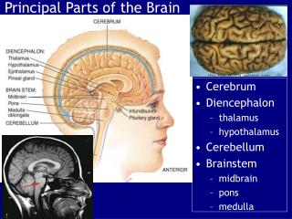
Autopsy Studies Show Structural Abnormalities in Autistic Brains • Short, dense, undeveloped brain cells, • Abnormalities observed primarily where MT levels are highest (amygdala, hippocampus, Purkinje cells, inferior olives, and pineal gland). Conclusion: Incomplete maturation of autistic brains may be due to low MT levels.
The Role of Metallothionein in the Development of Brain Cells • MT-3 assists in the pruning of brain cells, which makes space for growth of “new” cells, • MT-1 and MT-2 participate in the natural growth (development) of brain cells, • MT-3 is the primary agent for termination of growth of fully-developed brain cells.
Teamwork Between MT, GSH, Se“The Three Musketeers” • GSH is first line of defense against Hg, Pb, etc, but has limited capacity for toxic metals. • When > 10% of GSH is bound to toxic metals, additional toxics are transferred from GSH to MT. • Se increases kinetics of the GSH/MT antioxidant system by more than 50%. • For major exposures, most toxic metals depart the body bound to MT.
MT-Promotion Therapy • Formulation of 22 nutrients that promote genetic expression or functioning of MT, including Zinc, Glutathione, and Selenium, • Aimed at completion of brain maturation to enable gains in cognition, speech, and socialization, • Has resulted in higher frequency of autism recovery at Pfeiffer Treatment Center. U.S. Patent 7,232,575 (issued June, 2007)
Oxidative Damage Study 1 • Published in October, 2006. Archives of Neurology; Vol. 63:1161-1164. Authors Pratico, Walsh, McGinnis, and Yao. • Findings: Elevated oxidative damage to fats and vascular tissues for autistic subjects, compared to controls.
Oxidative Damage Study 2 • American Journal of Biotechnology and Biochemistry; 4(2):61-72, 2008. Authors: Evans, McGinnis, Walsh, Perry, Salomon, Lewis, et. al. • First direct evidence of oxidative damage inthe autistic brain. • Evidence of neurodegeneration in autism
Implications of Oxidative Damage Studies • Untreated autism may be neurodegenerative with oxidative damage causing slow, gradual loss of brain cells and IQ. • Antioxidant therapy may be necessary throughout the life of a person diagnosed with an autism spectrum disorder.
Clinical Evidence (n=7,000) of Neurodegeneration in Autism • Most young ASD patients appear quite bright • Many successfully treated children become mainstreamed and academic leaders, • Most adult autistics exhibit mental severe retardation.
Leukocyte Study “Altered Sulfur Amino Acid Metabolism in Immune Cells of Children Diagnosed with Autism”; J. Suh, W. Walsh, W. McGinnis, A. Lewis, and B. Ames. American Journal of Biochemistry & Biotechnology; 4 (2): 105-113, 2008.
Leukocyte Findings for ASD • SAMe levels 36% lower, • SAMe/SAH ratios 50% lower, • Homocysteine 180% higher, • Cysteine 40% lower, • GSH 25-60% lower.
Leukocyte Study Conclusion Evidence of increased inflammation, increased oxidative stress, and depressed immune function in autism.
Urine Pyrroles and Autism “Discerning the Mauve Factor, Part 1, 2.” Alternative Therapies in Health and Medicine, Vol. 14, No. 2, March, 2008. W.McGinnis, T.Audhya, W.Walsh, J.Jackson, J.McLaren-Howard, A.Lewis, P.Lauda, D.Bibus, F.Jurnak, R.Lietha, A.Hoffer. • 25-35% of ASD patients exhibit elevated pyrroles. • Urine HPL is a good marker for oxidative stress.
Comparison of Elemental Levels in Autism & Control Brains • Double blind, controlled study, • 176 brain tissues & 22 peripheral samples from U. of Maryland’s Autism Brain Bank, • Elemental analysis for 16 elements, including Hg, Pb, Cu, Zn, and Se using high-brilliance photons at ANL’s Advanced Photon Source), • First elemental assays ever attempted for autism & control brain tissues.
Brain Regions Studied • Cerebellum • Superior Cortex • Deep Cortex • White Matter Note: 20 autistic & 20 control tissue samples from each brain region
Summary of Findings • Abnormal levels of Ca, S, Fe, Zn in autism brains, • The abnormalities are strikingly different for male and female autistics, suggesting that male and female autism may have different genetic origins. • Mercury not detected (detection limit of about 100 ppb) Note: Article prepared for Neurology.
Distinctive Features of Autism • Strong genetic predisposition • Onset after environmental insult • High oxidative stress • Undermethylation • Incomplete brain maturation
Genetic Aspects of Autism • Strong genetic predisposition -- Higher concordance in siblings -- 60 to 80% concordance in identical twins • Influence of environmental factors -- Identical twin concordance not 100% -- Major differences in many identical twins.
QUESTION: How Can There Be An Epidemic of a Genetic Condition? ANSWER: The genetic defect involves a weakened ability to cope with environmental stresses
Timing of Environmental Insults is Important In Utero Autism evident at birth. Greater severity of symptoms. Mental retardation often present. After Birth Regressive autism. Symptoms depend on developmental stage during insult.
Severity of Environmental Insult Is Important Example:Disruption of key brain proteins during development of speech center. • Mild insultresults in speech delay. • Severe insult results in mutism.
Poly-Gene Nature of Autism • Current consensus that autism results from many genetic defects, rather than from a single gene. • A common factor in these genetic defects may be diminished ability to cope with oxidative stress.
What is Autism?Oxidative Stress Theory of Autism • Genetic tendency for depressed GSH, MT, Se, etc at intestinal and blood/brain barriers, • Inability to prevent Hg, Pb, Cd, and reactive oxygen specie from invading the brain. -- destruction of brain cells -- interruption of brain maturation process
Consequences of Oxidative Stress Mirror Classic Symptoms of Autism • Hypersensitivity to Hg and other toxic metals • Hypersensitivity to certain proteins (casein, gluten, etc) • Poor immune function • Disruption of the methylation cycle • Inflammation of the brain & G.I. tract. • Depletion of glutathione & metallothionein • Excessive amounts of “unbound” copper
Consequences of Oxidative Stress in the G.I. Tract • Destroys digestive enzymes needed to break down casein & gluten proteins, • Promotes candida/yeast levels, • Diminishes Zn levels and production of stomach acid, • Produces inflammation, • Ineffective barrier to toxic metals at the intestinal mucosa.
Most Popular Autism Therapies Enhance Antioxidant Protection • Chelation with DMSA, DMPS, EDTA, etc. • Methyl B-12 • Metallothionein Promotion • Transdermal or Injected Glutathione • Zn, Se, CoQ-10, Taurine, Vitamins A,C,D,E • Alpha Lipoic Acid • Risperdal
Mercury Questions • What % of autism cases are triggered by Hg? • Can “old” Hg stay in the brain and cause continuing damage? • How serious is the continuing daily exposure to Hg from the environment?
Chelation and Oxidative Stress • DMSA and DMPS are powerful antioxidants. • Chelation can provide antioxidant benefits even if toxic metals are not present. • For many patients, the primary benefits of chelation result from antioxidant properties, and not from removal of Hg or other metals. • Antioxidant benefits from chelation appear to “fade away” after about 2-4 weeks.
Primary Benefits of Chelation • Rapid removal of toxic metals from peripheral soft tissues & blood, thus preventing their access to the brain, • Powerful antioxidant
Limitations of Chelation • Does not fix intestinal or blood/brain barriers, rendering the patient vulnerable to future toxic exposures, • Antioxidant benefits are temporary, lasting only 2-4 weeks, • May not remove toxic metals from the brain, • Complicates Zn management.
Pfeiffer Treatment Protocol • Identification & individualized treatment of biochemical imbalances, • MT-Promotion therapy, • Selective use of adjunct therapies - CF/GF diet - Normalization of intestinal flora - Methylation therapies - Digestive enzymes - etc.