Urinary System Structures
Urinary System Structures. Figure 15.1a Organs of the urinary system. (a) Anterior view of urinary organs of a female (most unrelated abdominal organs have been removed). Rectum (cut). Uterus (part of female reproductive system). (a).

Urinary System Structures
E N D
Presentation Transcript
Figure 15.1a Organs of the urinary system. (a) Anterior view of urinary organs of a female (most unrelated abdominal organs have been removed). Rectum (cut) Uterus (part of female reproductive system) (a)
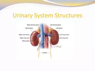
Figure 15.1a Organs of the urinary system. (a) Anterior view of urinary organs of a female (most unrelated abdominal organs have been removed). Hepatic veins (cut) Inferior vena cava Adrenal gland Renal artery Renal hilum Renal vein Aorta Kidney Ureter Iliac crest Rectum (cut) Uterus (part of female reproductive system) Urinary bladder Urethra (a)
Figure 15.2b Internal anatomy of the kidney. (b) Diagrammatic view of a coronally sectioned kidney, illustrating major blood vessels. (b)
Figure 15.2b Internal anatomy of the kidney. (b) Diagrammatic view of a coronally sectioned kidney, illustrating major blood vessels. Cortical radiate vein Cortical radiate artery Arcuate vein Renal column Arcuate artery Interlobar vein Interlobar artery Segmental arteries Renal cortex Renal artery Renal vein Minor calyx Renal pelvis Major calyx Renal pyramid Ureter Fibrous capsule (b)
Figure 15.3a Structure of the nephron. Cortical nephron Fibrous capsule Collecting duct Cortex Medulla Renal cortex Proximal convoluted tubule Renal pelvis Juxtamedullary nephron Glomerulus Ureter Distal convoluted tubule Loop of Henle Renal medulla (a)
Figure 15.3b Structure of the nephron. (b) Detailed anatomy of a nephron and its associated blood supply. (b)
Figure 15.3b Structure of the nephron. (b) Detailed anatomy of a nephron and its associated blood supply. Peritubular capillaries Proximal convoluted tubule (PCT) Glomerular capillaries Glomerular (Bowman’s) capsule Distal convoluted tubule (DCT) Efferent arteriole Afferent arteriole Cells of the juxtaglomerular apparatus Cortical radiate artery Arcuate artery Arcuate vein Cortical radiate vein Collecting duct Loop of Henle (b)
Figure 15.4 The kidney depicted schematically as a single large, uncoiled nephron. KEY: 1 a 2 b c 3 Urine
Figure 15.4 The kidney depicted schematically as a single large, uncoiled nephron. Afferent arterioles Glomerular capillaries KEY: Efferent arterioles Cortical radiate arteries Glomerular Filtration: Water and solutes smaller than proteins are forced through the capillary walls and pores of the glomerular capsule into the renal tubule. 1 Glomerular capsule a Rest of renal tubule Tubular Reabsorption: Water, glucose, amino acids, and needed ions are transported out of the filtrate into the tubule cells and then enter the capillary blood. 2 Peritubular capillaries b c Tubular Secretion: H+, K+, creatinine, and drugs are removed from the peritubular blood and secreted by the tubule cells into the filtrate. To cortical radiate veins 3 Urine