Download
1 / 13
250 likes | 913 Vues
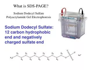
What is SDS-PAGE?. Sodium Dodecyl Sulfate Polyacrylamide Gel Electrophoresis. Sodium Dodecyl Sulfate: 12 carbon hydrophobic end and negatively charged sulfate end. SDS PAGE is a technique for separating proteins based on their molecular weight.

E N D
What is SDS-PAGE? Sodium Dodecyl Sulfate Polyacrylamide Gel Electrophoresis Sodium Dodecyl Sulfate: 12 carbon hydrophobic end and negatively charged sulfate end
SDS PAGE is a technique for separating proteins based on their molecular weight. Large proteins migrate slowly through the gel matrix and small proteins migrate quickly through the matrix The porous gel matrix is composed of the polymer polyacrylamide
Link Cross Link polymerization The polymerization reaction is catalyzed by TEMED (tetramethyl Ethylenediamine and APS (ammonium persulfate)
Pore size is determined by: The percentage of acrylamide used. Concentrations range from 5% to 30% Acrylamide Concentration Affects Protein Mobility
- - - - - - - - - - - - - - - - - - - - - - - - - - - - - SDS - - - - - - - - - - - - - - - - - - - - - - - - - - - - - The hydrophobic region of SDS binds to the hydrophobic region of proteins and unfolds them. This also covers the proteins with a negative charge. All proteins now have a net negative charge.
- - - - S S - - - S S S - - - - - - - - - - - - - - - - - - - - - - - - - - - - - - - - - - - - - - - - - - - - - - - - - - S SDS - - - - - - - - - - - - - - - - - - - - - - - - - - - - - - - - - - - - - - - - - - - - - - - - - - - - - - - Disulfide bonds must be broken to completely unfold the protein. Reducing agents/antioxidents are used to break the disulfide bonds. DTT: Dithiothreitol and BME: Beta mercaptoethanol SDS Plus DTT
Negatively charged protein samples are loaded into wells at the top of the gel An electric current is applied so that there is a positive charge at the bottom of the gel and a negative charge at the top of the gel. The negatively charged proteins will migrate to the bottom of the gel according to size.
Visualizing the proteins: • Types of Stains: • Coomassie Blue – Sensitivity of 0.1ug protein per band • Traditional method requires staining followed by destaining to remove background gel staining. • GelCode Blue from Pierce does NOT stain the gel and requires no destaining. Increased sensitivity of 25ng protein per band • 2. Silver Stain – Sensitivity of 2ng protein per band • Glassware and plasticware must be very clean to prevent stain from precipitating out of solution. • Prone to high background due to impurities in the acrylamide and surface artifacts such as fingerprints
Lane 1. Kaleidoscope Markers 2. Shark 3. Salmon 4. Trout 5. Catfish 6. Sturgeon 7. Actin and Myosin Standard
Comparison of the sensitivity of SDS PAGE stains: Identical SDS-polyacrylamide gels were stained with A) SYPRO Orange protein gel stain B) SYPRO Red protein gel stain C) silver stain D) Coomassie brilliant blue stain
Assemble gel box and make buffers • 2. Mix sample, reducing agent and loading buffer • 65ul sample • 10ul reducing agent • 25ul loading buffer • 3. Boil 5 minutes • 4. Centrifuge 5 sec • 5. Load gel, 75ul for each sample Lane Sample 1 Flow Through Fraction 2 Flow Through Fraction 3 Flow Through Fraction (good column) 4 Elution Fraction 5 Elution Fraction 6 Elution Fraction (good column) 7 Protein ladder 8 Unpurified HSA-Unconcentrated 9 Unpurified HSA-Concentrated 10 Loading buffer Load the protein ladder asymmetrically for easier identification of samples
6. Run at 200V for approximately 1 hour • 7. Remove gel and wash 3 times with water, 5 minutes each • Incubate gel in fixative for 15 minutes • Wash 3 times with water, 5 minutes each • Incubate in GelCode Blue for approximately 1 hour • Rinse gel with water