Laboratory Manual for Diagnosing Bacterial Conjunctivitis via Conjunctival Discharge
This comprehensive manual outlines the procedures for diagnosing bacterial conjunctivitis through the examination and culture of conjunctival discharge. Key aspects include specimen collection, processing methods, rejection criteria, and factors affecting test results. Emphasis is placed on immediate processing due to lysosomes in tears that can kill the organism. Techniques for identification and susceptibility testing of isolated bacteria are discussed, along with pre- and post-specimen processing steps. Suitable for medical microbiology laboratories and healthcare professionals.

Laboratory Manual for Diagnosing Bacterial Conjunctivitis via Conjunctival Discharge
E N D
Presentation Transcript
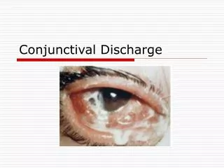
بسم الله الرحمن الرحيم ConjunctivalDischarge 2013-2014 Diagnostic Medical Microbiology-Laboratory Manual
Conjunctival Discharge Aim of the test An etiological diagnosis of bacterial conjunctivitis by aerobic cultivation with identification and susceptibility test of the isolated bacteria. Types of specimen Two swabs from discharge from the eye(s). Criteria of specimen rejection Inappropriate specimen transport device; mislabeled specimen; unlabeled specimen; specimen received after prolonged delay (usually more than two hour); specimen received in expired transport media.
Pre specimen processing Who is authorized to order the test Physician. Quantity of specimen Sufficient amount on swab. Time relapse before processing the sample Eye specimen should be processed immediately because tears contains lysosomes which may kill the organism. Storage Maintain specimen swab at room temperature .Don’t refrigerate.
Pre specimen processing Conjunctival discharge Specimen collection:- Pull down the lower eyelid so that the lower conjunctival fornix is exposed. Swab the fornix without touching the rim of the eyelid with the sterile cotton swab. Place the swab immediately in a bacterial transport medium or, if the specimen is brought to the laboratory immediately, in a sterile test tube with 0.5 mL of phosphate buffered saline.
Specimen processing for conjunctiva • Direct Visual Examination • All material submitted for culture should always be smeared and examined directly by gram stain or other appropriate techniques. • Specimen in which chlamydia is suspected can be stained immediately with monoclonal antibody conjugated to fluorescein for detection of elementary bodies or inclusions. • Culture • Because the constant washing action of the tears the number of organisms recovered from cultures of certain eye infection may be relatively low, so Conjunctival scrapings place directly onto media yield the best results.
Specimen processing for conjunctiva • One should use blood, MacConkey and chocolate agar plates, because potential pathogen may present in an eye without causing infection it maybe very helpful when any one eye is infected to culture both eyes, If a potential pathogen grows in cultures of both infected and un infected eye the organisms may not be causing the infection, now ever if the organism only grows in culture from the infected eye, it is most likely the causative. • Non Culture Methods • ELISA and DIFA staining are now available for detection of Chlamydia trachomatis
Direct Immuno-fluorescence (DIF) Antibody labeling for Chlamydia trachomatis
Chlamydia trachomatis With Giema stain With Iodine stain Giemsa stain of Chlamydia inclusion bodies (purple "caps" on epithelial cell).
Post specimen processing • Interfering factors: • Patient on antibiotic therapy. • Improper sample collection. • Result reporting: • Report Gram stain finding as an initial report. • Report the isolated pathogen and its sensitivity pattern as a final report. • Turn around time: • Gram stain result should be available half hour after specimen receipt. • Isolation of a possible pathogen can be expected after 2-3 days. • Negative culture will be reported out 1-2 days after the receipt of the specimen.
END LECTURE Any Questions ?