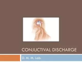
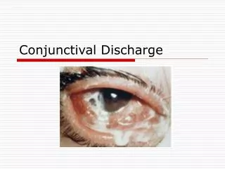
Conjunctival Discharge
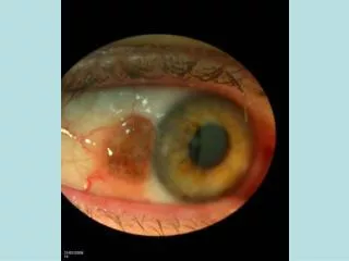
Conjunctival Discharge. Conjunctivitis. Inflammation Erythema Several causes: Bacterial Viral Allergic Chemical. Conjunctivitis - Discharge. Discharge Cause Purulent Bacteria Clear Viral White, stringy mucous Allergies. Bacterial conjunctivitis. Purulent discharge

Conjunctival Discharge
E N D
Presentation Transcript
Conjunctivitis Inflammation Erythema Several causes: Bacterial Viral Allergic Chemical
Conjunctivitis - Discharge DischargeCause Purulent Bacteria Clear Viral White, stringy mucous Allergies
Bacterial conjunctivitis Purulent discharge Conjunctival hyperemia
Viral Conjunctivitis Adenovirus Systemic viral infections Painful Herpetic Discordant lack of pain
Viral conjunctivitis Diffuse redness Watery discharge
Aim of the test • An etiological diagnosis of bacterial conjunctivitis by aerobic cultivation with identification and susceptibility test of the isolated bacteria . • Types of specimen • Discharge from the eye(s).
Specimen collection • Pull down the lower eyelid so that the lower conjunctival fornix is exposed. • Swab the fornix without touching the rim of the eyelid with the sterile cotton swab. • Place the swab immediately in a bacterial transport medium or, the specimen is brought to the laboratory immediately, in a sterile test tube with 0.5 mL of buffered saline (pH 7). • Take Sufficient amount on the swab
Time relapse before processing the sample • Eye specimen should be processed immediately because tears contains lysosomes which may kill the organism
Media • Blood Agar • Chocolate Agar • MacConkey Agar • Fluid Thioglycollat
Ear Discharge • Aim of the test • Aetiological diagnosis of otitis external or otitis media by aerobic and anaerobic culture with identification and susceptibility test of the isolated organism (s). • Types of specimen • Pus from the external or middle ear.
Specimen collection • Collect a specimen of the discharge on a thin, sterile cotton wool or Dacron swab. • Place the swab in a container with the transport medium, breaking off the swab stick to allow the stopper to be replaced tightly. • Label the specimen and send it to the laboratory. • Time relapse before processing the sample Not more than 2 hours
Media • Blood Agar, • Chocolate Agar, • MacConkey Aga • Fluid thioglycolla
Background & Terminology • Vaginitis : significant inflammatory response in vaginal wall. Accompanied by high number of leukocytes in vaginal fluid. Found with candida and trichomonas infections. • Vaginosis: minimal inflammatory response with few leukocytes in vaginal wall. Associated with increase in bacterial concentrations. • Leukorrhoea : a non-infective, non-bloodstained physiological vaginal discharge.
Clinical approach • Physical Exam : • Appearance of discharge. • Erythema and edema of vaginal mucosa • pH levels • Diagnostic Tools: • pH : Nitrazine paper • Wet prep: microscopic examination of discharge • KOH prep: dissolves cellular debris leaving pseudohyphae of candida. • Whiff test: Fishy odor of BV • Culture
Common Causes • Normal discharge (30%) • Bacterial Vaginosis (23-50%) • Candida Vulvovaginitis (20-25%) • Trichomonas vaginitis (5-15%) • Mixed infection or Sexually Transmitted Disease (20%)
Whiff Test The vaginal discharge of patients with BV has a characteristic fishy odor due to increased activity of anaerobic species. Addition of KOH will augment this odor.
Aim of the test • Isolate and identify potentially aerobic pathogenic organisms including • Gardnerella vaginalis and group B Streptococcus; establish the diagnosis of gonorrhea, medical/legal cases.
Types of specimen • Swab of vagina, cervix, discharge, aspirated endocervical, endometrial, prostatic fluid, or urethral discharge. • Use swab to inoculate Jembec for transport to the laboratory and recovery of Neisseria gonorrhoeae; swab • should also be sent in transport device.
Specimen collection • Do not use lubricant on speculum. • Cervical mucous should be removed first before inserting swab into endocervical canal, move swab from side to side allowing several seconds for absorption of organisms by the swab. • Return swab to the transport tube and label.