Download
1 / 24
270 likes | 632 Vues
Dep. of Chemistry & Biochemistry Prof. Indig. Chemistry 501 Handout 4 The Three-Dimensional Structure of Proteins Chapter 4. Lehninger. Principles of Biochemistry. by Nelson and Cox, 5 th Edition; W.H. Freeman and Company. A protein’s conformation is stabilized

E N D
Dep. of Chemistry & Biochemistry Prof. Indig Chemistry 501 Handout 4The Three-Dimensional Structure of ProteinsChapter 4 Lehninger. Principles of Biochemistry. by Nelson and Cox, 5th Edition; W.H. Freeman and Company
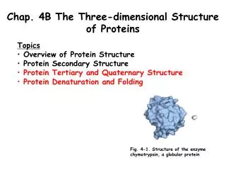
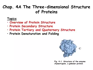
A protein’s conformation is stabilized largely by weak interactions Proteins in any of their functional, folded conformations are called native proteins. Glycine Quaternary structure of deoxyhemoglobin (a) A ribbon representation (b)A space-filling model Structure of the enzyme chymotrypsin, a globular protein (PDB file 6GCH) The known 3-D structures of proteins are archived in the Protein Data Bank (PDB). Each structure is assigned a four-character identifier, or PDB ID.
Protein Architecture - Primary Structure The planar peptide bond Each peptide bond has some double bond character due to resonance and cannot rotate Series of rigid planes sharing a common point of rotation at Ca Three bonds separate the a carbons in a polypeptide chain = = 180o when the peptide is in its fully extended conformation and all peptide groups are in the same plane
Convention: In this conformation,f = y = 0o In a protein, this conformation is prohibited by steric overlap between an a-carbonyl oxygen and an a-amino hydrogen atom Ramachandran plot for L-Ala residues Conformations deemed possible are those that involve little or no steric interference, based on calculations using known van der Walls Radii and bond angles
Protein Architecture - Secondary Structure A few types of secondary structures are particularly stable and occur widely in proteins. Most prominent: a helix and b conformations. Space-filling model Note: The a helix is not hollow Ball-and-stick model Ball-and-stick model of a right-handed a helix, showing the intrachain H bonds The a helix as viewed from one end, looking down the longitudinal axis The atoms in the center of the a helix are in very close contact Helical wheel representation of an a helix.
Not all polypeptides can form a stable a helix. Interactions between side chains can stabilize of destabilize this structure The identity of the amino acid residues near the ends of the a-helical segment also affects the stability of the helix Arg103 Asp100 Interaction between R groups of amino acids three residues apart in an a helix Troponin C (shown: a helix segment 13 residues long) The four amino acid residues at each end of the helix do not participate fully in the helix hydrogen bonds
b turns are common in proteins The b conformation organizes polypeptide chains into sheets 180o turn involving four amino acid residues Gly and Pro residues often occur in b turns Gly - small and flexible Pro - the cis configuration is particularly amenable to a tight turn b sheets
Common secondary structures have characteristic bond angles and amino acid content Ramachandran plots for a variety of structures Common secondary structures can be assessed by circular dichroism (CD) spectroscopy Pyruvate kinase (all amino acid residues except Gly)
Protein tertiary and quaternary structures a helix loop deoxyhemoglobin b conformation Tertiary structure includes long-range aspects of amino acid sequence Quaternary structure includes the three-dimensional arrangement of polypeptide chains in multisubunit proteins
Example of quaternary structure Fibrous Proteins (structure, support, shape, protection) - Polypeptide chains arranged in long strands of sheets left-handed supertwisted coiled coil Disulfide (-S-S-) bonds stabilize the quarternary structure Permanent waving a-Keratin
Collagen (connective tissue, tendons, cartilage, organic matrix of bone, cornea) Structure of collagen fibrils The three-stranded collagen superhelix shown from one end (ball-and-stick representation) Three helix wrap around one another with a right-handed twist • chain: repeating tripeptide sequence (generally Gly-X-Y, where X is often • Pro and Y is often 4-Hyp)adopts a left-handed helical structure with three residues per turn
Structure of silk Fibroin : layers of antiparallel b sheets rich in Ala and Gly residues Permits close packing and interlocking of R groups Sheets held together by numerous weak interactions, rather than covalent bonds such as disulfide bonds in a-keratin Strands of fibroin (blue) emerge from the spinnerets of a spider (colorized electron micrograph)
Globular Proteins Structures compact and varied Tertiary structure of a small globular protein:sperm whale myoglobin e.g. human serum albumin 585 residues in a single chain (Mr 64,500) Approximate dimensions its single polypeptide chain would have if it occurred entirely in: a) Polypeptide backbone shown in a ribbon representation b) Surface contour image: useful for visualizing pockets in the protein c) Ribbon representation including side chains for the hydrophobic residues Leu, Ile, Val, and Phe d) Space-filling model with all amino acid side chains The heme group
Supersecondary structures (motifs, or simply folds) Particularly stable arrangements of several elements of secondary structure and the connections between them. Motifs
Protein motifs are the basis for protein structural classification Constructing large motifs from smaller ones The Structural Classification of Proteins (SCOP) database. Protein structures divided into four classes: all a all b a/b (a and b segments interspersed or alternate) a+b (a and b regions are somewhat segregated) Within each class: tens to hundreds of different folding arrangements, built up form increasingly identifiable structures.
Protein quaternary structures range from simple dimers to large complexes Viral capsids Quaternary structure of deoxyhemoglobin a) The coat protein of poliovirus assemble into an icosahedron 300 Angstrons in diameter b) Tabaco mosaic virus: rod-shaped virus 3,000 Angstrons long and 180 Angstrons in diameter with helical symmetry
Protein denaturation and folding Loss of protein structure results in loss of function
The thermodynamics of protein folding depicted as a free-energy funnel Polypeptides fold rapidly by a stepwise process
Folding for many proteins is facilitated by the action of specialized proteins (chaperones) E. Coli chaperone proteins DnaK and DnaJ
Chaperonins in protein folding GroEL/GroES complex Proposed pathway for the action of the E. coli chaperonins GroEL and GroES.
Defects in protein folding may be the molecular basis for a wide range of genetic disorders Formation of disease-causing amyloid fibrils
Prion Diseases Stained section of cerebral cortex from autopsy of a patient with Creutzfeldt-Jakob disease shows spongiform (vacuolar) degeneration, the most characteristic neurohistological feature. Structure of the globular domain of human PrP in monomeric (left) and dimeric (right) forms. Proteinaceousinfectiousonlyprotein (PrP)