Download
1 / 46
670 likes | 1.4k Vues
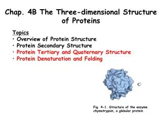
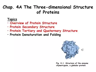
Chapter 4 The Three-Dimensional Structure of Proteins. 4.1 Overview of Protein Structure. Protein Conformation. Conformation Spatial arrangement of atoms in a protein Tendency to have the lowest Gibbs free energy (highest stability) Noncovalent interactions determining protein conformation

E N D
Chapter 4 The Three-Dimensional Structure of Proteins
Protein Conformation • Conformation • Spatial arrangement of atoms in a protein • Tendency to have the lowest Gibbs free energy (highest stability) • Noncovalent interactions determining protein conformation • Maximum hydrogen bonding within the protein • DH for H bonds in protein ≈ DH for H bonds with water • DS > 0by H bonding in protein caused by decrease in solvation shell of structured water • Hydrophobic interaction • Hydrophobic residues are buried in the protein interior • Ionic interactions (salt bridge) • Disulfide bonds • Native proteins • Proteins in any of their functional, folded conformation
The Peptide Bond is Rigid and Planar • Double bond character of peptide bond • Resonance between the carbonyl oxygen and the amide nitrogen • 6 atoms of the peptide group lie in a single plane • No free rotation of peptide C-N bond (trans) • Rotation of peptide chain • f: rotation angle of N-Ca • y : rotation angle of Ca-C f, y = 180 (or -180)
Ramachandran Plot • Rotation of peptide chain • -180 <f &y < 180 • f, y = 0 • Reference point for describing the angels of rotation • Two peptide bonds are in the same plane • Restricted by steric overlap • Ramachandran Plot • Plotting of the allowed values of fvs.y
4.2 Protein Secondary Structure Secondary structure • Local conformation of polypeptide • helix, sheet : 60% of the polypeptide chain • Random coils and - turn
Helix • Hydrogen bond between carbonyl O (n) and amid H (n+4) • Right-handed helix • One turn: 5.4 Å along the axis, 3.6 amino acids • y = -45 to -50 f= -60 • Side chains point outward
Amino Acid Sequence Affects a Helix Stability • Amino acids destabilizing a helix • Electrostatic repulsion • Glu, Lys, Arg • Bulkiness & shape of adjacent R groups • Asn, Ser, Thr, Cys • Restricted rotation • Pro • No N-Ca (f)rotation kink • No H in N for hydrogen bonding • Flexible rotation • Gly • Tendency to form coil structure different from a helix
Amino Acid Sequence Affects a Helix Stability • Interaction between amino acid residues at the ends of the helical segment and the electric dipole of a helix • (+) charged a.a near C-terminus • (-) charged a.a near N-terminus
Constraints for the stability of a-helix • Intrinsic propensity of a.a to form a-helix • Interactions between side chains • Bulkiness of adjacent side chains • Occurrence of Pro and Gly residues • Interactions between a. a at the ends of the helical segment and the electric dipole inherent to the a-helix
b Conformation • b stand • Zigzag polypeptide backbone • b sheet, b-pleated sheet • Hydrogen bonding between adjacent b strands • Parallel • Antiparallel • Amino acids for specific b sheet structure • Stacking of b sheet • b-keratins (silk fibroin, spider web) • Rich in small amino acids: Gly, Ala
b Turns • Connecting elements • 1/3 of amino acids in a globular protein • Turns and loops • b turns • Connecting the ends of two adjacent segments of antiparallel b sheet • 180o turns involving 4 amino acids and hydrogen bonding • Gly : small and flexible • Pro : cis configuration amenable to a tight turn
Bond Angles of Amino Acid Content of Secondary Structure • Relatively restricted range of y and fdepending on the types of secondary structure • Different distribution of amino acids in different secondary structures
4.3 Protein Tertiary and Quaternary Structure
Higher Protein Structure • Tertiary structure • Overall 3D arrangement of all atoms in a protein • Quaternary structure • Arrangement of protein subunits • Classification by higher structure • Fibrous proteins • Single type of secondary structure • Provide support, shape, and external protection • Globular proteins • Several types of secondary structure • Enzymes and regulatory proteins
Fibrous Proteins • Characteristics of fibrous proteins • Strength and flexibility • Water insoluble • High concentration of hydrophobic amino acids
a keratin • Structural protein for hair, wool, feathers, nails, hooves, horns • Providing strength • Coiled coil (left handed twist) of a-helix with hydrophobic amino acids (A, I, V, M, F) • Forming fibers by hydrophobic interactions
Disulfide bonds • The more S-S bonds the harder the structure • Permanent wave • Reducing of disulfide bond Generation of new disulfide bond
Collagen • Providing strength in connective tissue • Tendon, cartilage, organic matrix of bone, cornea • Structure • Left-handed helix with 3 a.a./turn : a chain • Right-handed supertwist of 3 a chains • Amino acid composition • Repeating tripeptide unit, Gly-X, Y • X; Pro, Y; 4-Hyp • 35% Gly, 11% Ala, 21% Pro and 4-Hyp • Gly is essential for the structure • Mutation genetic disease • Very low nutritional value • Very close packing • Collagen fibrils • Crosslinking of collagen molecules by involving lysine, hydroxylysine, histidine
Silk Fibroin • Produced by insects and spiders • b conformation • Rich in Ala and Gly • Close packing of b-sheets and interlocking alignment of R groups • Stabilization by hydrogen bonding and van der Waals interactions • Flexible Strand of fibroin emerging from the spinnerets of a spider
Globular Proteins • Globular proteins • Compact • Structural diversity to carry out diverse functions • Myoglobin • Structure determined by x-ray diffraction studies (John Kendrew, 1950’s) • Oxygen carrier in muscle : containing heme group • 153 a.a
Diverse Tertiary Structure of Globular Proteins • Shared properties w/ myoglobin • Compact folding • Hydrophobic side chains in the interior • Hydrophilic sided chains on the surface • Stabilization by non-covalent interactions
Common Structural Patterns • Motifs (folds or supersecondary structures); folding pattern • Stable arrangements of several elements of secondary structure • Domains • Stable, globular units; distinct functions
Rules of common protein folding patterns • Rules of common protein folding pattern • Hydrophobic interaction • Burial of hydrophobic R groups • Layers of 2nd structures; b-a-b loop, a-a corner • In general, a helices and b sheets are in different structural layers • Stacking of the adjacent polypeptide segments • No crossover connection • b conformation is most stable with slight right-handed twist
Classification of Protein Structures • Structural classification of proteins (SCOP) database • Classification • All a • All b • a/b : a and b segments are interspersed or alternate • a + b : a and b regions are segregated • < 1,000 different folds or motifs • Protein family • Proteins with similarities in • Primary sequence • (and/or) Structure • Function • Superfamily • Families with little primary sequence similarity but with similarities in motifs and function • Tracing structural motifs using protein database • Useful to identify evolutionary relationships (protein 3rd structure is more conserved than A. a. sequence)
Quaternary Structure • Hemoglobin • Tetramer : two a chains and two b chains • Dimer of ab protomer • Symmetric patterns of multimeric proteins with identical subunits • Rotational symmetry • Cyclic symmetry • Single axis for rotation : Cn , n fold rotation axis • Dihedral symmetry • Intersecting twofold rotational axis and n fold axis at right angles : Dn, 2n protomers • Icosahedral symmetry • 12-cornered polyhedron with 20 equilateral triangular faces • Virus coats and capsids • Helical symmetry • Capsid of tobacco mosaic virus • Actin filaments
Symmetric patterns of multimeric proteins Helical symmetry
Protein Denaturation • Denaturation • A loss of three-dimensional structure sufficient to cause loss of function • Not necessarily means complete unfolding or random conformations • Abrupt unfolding over a narrow temperature range • Cooperative unfolding process • Denaturing agents • Heat • Affect weak interactions (H bonds) • pH • Alternation of the protein net charge • Electrostatic repulsion, disruption of H bonds • Organic solvents (alcohol, acetone), urea, guanidine HCl, detergents • Disruption of hydrophobic interactions
Amino Acid Sequenc Determines Tertiary Structure • Renaturation • Reversal of denaturation • Amino acid sequence contains all the information required to protein folding • First experimental evidence by Christian Anfinsen (1950s) • Denaturation of ribonuclease with urea and reducing agent • Spontaneous refolding to an active form upon removal of the denaturing reagents
Protein Folding • Protein folding in living cells • Not a random, trial-and-error process • E. coli : make 100 a.a. protein in 5 sec • 10 possible conformations/ a.a. 10100 conformations • 10-13 sec for each conformation 1077 years to test all the conformations “Levinthal’s Paradox”
Models for Protein Folding • Hierarchical folding • From local folding (a helix, b sheets) to entire protein folding • Molten globule state model (hydrophobic collapse) • Initiation of folding by spontaneous collapse by hydrophobic interactions
Thermodynamics of Protein Folding • Free-energy funnel • Unfolded states • High entropy and high free energy • Folding process • Decrease in the number of conformational species (entropy) and free energy • Semistable folding intermediates
Molecular Chaperones • Molecular chaperones • Proteins facilitating protein folding byinteracting with partially or improperly folded proteins • Classes of molecular chaperones • Hsp70 • Induced in stressed cells (heat shock protein) • Binding to hydrophobic regions of unfolded proteins, preventing aggregation • Cyclic binding and release of proteins by ATP hydrolysis and cooperation with co-chaperones (Hsp40 etc.) • E. coli: DnaK (Hsp70), DnaJ (Hsp40) • Chaperonin • Protein complex providing microenvironments for protein folding • E. coli : 10~15% protein require GroES (lid) and GroEL • Isomerases in protein folding • Protein disulfide isomerase (PDI) • Shuffling disulfide bonds • Peptide prolyl cis-trans isomerase (PPI) • Interconversion of the cis and trans isomers of Pro peptide bonds
Protein Folding and Diseases • Cystic fibrosis • Misfolding of cystic fibrosis transmembrane conductance regulator (CFTR; Cl- channel) • Neurodegenerative diseases • Alzheimer’s, Parkinson’s, Huntinton’s desease, ALS • Prion diseases • Mad cow disease (bovine spongiform encephalopathy, BSE) • Kuru, Creutsfeldt-Jakob disease in human • Scrapie in sheep • Prion : proteinaceous infectious only protein • PrPSc (scrapie) prion form converts PrPC to PrPSc
Neurodegenerative disorders • The amyloid formation is the common phenomenon observed in various neurodegenerative disorders, including Parkinson’s disease, Alzheimer’s disease, Huntington’s chorea, Amyotrophic lateral sclerosis, Prion disease, etc. Parkinson’s disease Alzheimer’s disease Prion disease [mad cow disease]
degradation Misfolded protein Folded state Amino acids refolding Accumulation Amyloid formation Degenerative disorders
Creutzfeldt-Jakob disease [Spongiform encephalopathies]