The Human Eye
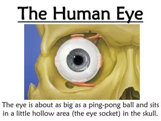
The Human Eye. Optics. VISION. Sclera The sclera is the white of the eye. "Don't shoot until you see their scleras." Exterior is smooth and white Interior is brown and grooved Extremely durable Flexibility adds strength Continuous with sheath of optic nerve Tendons attached to it.


The Human Eye
E N D
Presentation Transcript
The Human Eye Optics
Sclera The sclera is the white of the eye. "Don't shoot until you see their scleras." • Exterior is smooth and white • Interior is brown and grooved • Extremely durable • Flexibility adds strength • Continuous with sheath of optic nerve • Tendons attached to it Eye Parts and their function:
Sclera • The sclera is the white of the eye. • Exterior is smooth and white • Interior is brown and grooved • Extremely durable • Flexibility adds strength • Continuous with sheath of optic nerve • Tendons attached to it
The Cornea • The cornea is the clear bulging surface in front of the eye. It is the main refractive surface of the eye. • Primary refractive surface of the eye • Normally transparent and uniformly thick • Richly supplied with nerve fibers • Sensitive to foreign bodies, cold air, chemical irritation
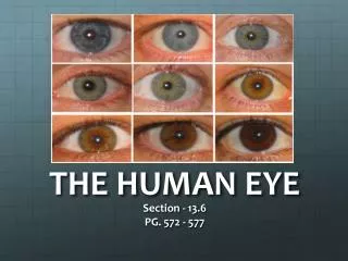
Iris/Pupil • Iris is heavily pigmented • Sphincter muscle to constrict or dilate the pupil • Pupil is the hole through which light passes • Eye colour(brown, green, blue, etc.) dependent on amount and distribution of the pigment melanin
Lens • Transparent body enclosed in an elastic capsule • Made up of proteins and water • Consists of layers, like an onion, with firm nucleus, soft cortex • Young person can change shape of the lens via ciliary muscles • Contraction of muscle causes lens to bulge • At roughly age 50, the lens can no longer change shape • Becomes more yellow with age: Cataracts
Vitreous Humor • Fills the space between lens and retina • Transparent gelatinous body • Also maintains eye shape
Retina • The retina is the film of the eye. • It converts light rays into electrical signals and sends them to the brain through the optic nerve. • The sides of the retina are responsible for our peripheral vision. • The center area, called the macula, is used for our fine central vision and color vision. • The retina is where most the problems leading to vision loss occur
3 leading causes of blindness retina damage: 1. Retinitis Pigmentosa 2. Macular Degeneration 3. Diabetic Retinopathy
Photoreceptors • The retina is composed of two types of photoreceptor cells. When light falls on one of these cells, it causes a chemical reaction that sends an electrical signal to the brain. 2 types: • Cone Cells • Rod Cells
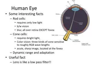
Cone cells • give us our detailed colourdaytime vision • there are 6 million of them in each human eye
Rod cells • are about 500 times more sensitive to light than cone cells; they give us our dim light or night vision • There are 120 million rod cells in the human eye
Optic nerve • Each optic nerve has about 1.2 million nerve fibers • This is the cable connecting the eye to the brain
Cilliary Muscle • Changes the shape of the lens • It relaxes to flatten the lens for distance vision; for close work it contracts rounding out the lens
Handout • Label the Diagram • Complete the table – structure and function
Here is a demonstration of the natural permanent scotoma (blind sopt): Close your left eye. Fixate on the cross with your right eye. This will cause the image of the cross to fall on your fovea. Adjust the viewing distance until the black spot disappears. When this happens, the image of the spot is falling on your blind spot. What do you see (or not see) when you do this with the top figure? What happens when the gap in the bottom figure falls on your blind spot?
Results • You should see the "smiley" in the top figure disappear when it falls in your blind spot. • When the gap in the bottom figure falls on the blind spot, the visual system "fills in" the line. So why don't we notice the blind spot in normal vision? 1. We have two eyes and the blind spots are in non-corresponding locations (they are nasally located (towards the nose) on the retina so the blind spots are temporal (towards the temple) in the visual field). 2. In addition, the filling in process makes the blind spot less noticeable especially in a peripheral area of sight that has less visual acuity (the ability to see detail).