The Human Eye
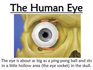
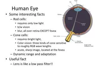
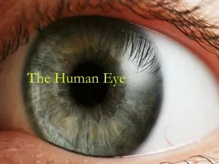
The Human Eye. How the eye works. Light rays enter the eye through the clear cornea, pupil and lens. These light rays are focused directly onto the retina, the light-sensitive tissue lining the back of the eye.


The Human Eye
E N D
Presentation Transcript
How the eye works • Light rays enter the eye through the clear cornea, pupil and lens. • These light rays are focused directly onto the retina, the light-sensitive tissue lining the back of the eye. • The retina converts light rays into impulses, sent through the optic nerve to your brain, where they are recognized as images. • 70% of the eye's focusing power comes from the cornea and 30% from the lens.
The Retina • The retina is composed of 2 different types of cells that sense different things • Rods are highly light sensitive (concentrated around the outside of retina) • Cones are colour sensitive (concentrated in the centre of retina)
Eye Accomodation • Accomodation is the changing of the shape of the lens of the eye by eye muscles to produce a sharply focused image on the retina • Some vision problems are caused by poor accomodation of the eyes or refractive errors • 4 common types of refractive errors occur
1. Myopia • In myopia (nearsightedness), the distance between the cornea and the retina is too long or the power of the cornea and the lens is too strong. • Light rays focus in front of the retina instead of on it. • Close objects will look clear, but distant objects appear blurry. Myopia, or nearsightedness
2. Hyperopia • In hyperopia (farsightedness), the distance between the cornea and the retina is too short. • Light rays are focused behind the retina instead of on it. • Distant objects will look clear, but close objects will appear blurred. Hyperopia, or farsightedness
3. Presbyopia • Occurs as people age… • The lens loses its elasticity the ability to see things up close is lost - the lens can no longer change shape. • Also corrected using converging lenses for reading glasses • (When we are young, the lens in our eyes is flexible and is able to change focus easily between near and far objects, like an autofocus on a camera.) • (At around age 40, this flexibility begins to gradually decrease, making it more difficult to see objects up close, unless the eye has nearsightedness. )
4. Astigmatism • In astigmatism, the cornea is curved unevenly—shaped more like a football than a basketball. • Light passing through the uneven cornea is focused in two or more locations. • Distant and close objects may appear blurry. • Corrected using eyeglasses, contact lenses that reshape the cornea or laser surgery to reshape the cornea Astigmatism occurs when light passes through football-shaped cornea and/or lens
How to test for Astigmatism • 1. If you have contacts or glasses, wear them. 2. Sit about 14 inches away from your computer screen. 3. Cover one eye.4. Note how the lines and squares appear (for example, wavy or blurred). 5. Test the other eye in the same manner.
What is refractive surgery? • A group of outpatient surgical procedures used to alter how your eye focuses light rays on the retina, thereby improving vision and reducing dependence on glasses and contact lenses. • In most cases, refractive surgery affects the shape of your cornea to redirect how light is focused onto the retina. Popular procedures include LASIK, LASEK, PRK and CK. Refractive surgery procedure on the cornea
What is refractive surgery? • Most refractive surgery is performed on the cornea and affects only the front of your eye, while the rest of your eye will change naturally as you age. • In some cases, refractive surgery procedures don’t reshape the cornea; instead, the eye’s natural lens is either replaced or enhanced by an implantable lens that helps correct vision.
What is Refractive Lens Exchange (RLE)? • A non-laser procedure where the natural, non-cataractous, lens of the eye is removed and replaced with an artificial, intraocular lens (IOL). • The cornea is not reshaped. • Used to treat moderate to high degrees of nearsightedness (myopia), farsightedness (hyperopia) and patients who are not LASIK candidates. Typical intraocular lenses (IOLs) used in the refractive lens exchange (RLE) procedure
How is the RLE procedure performed? • The IOL is implanted in a surgical procedure and performed on an outpatient basis under local or topical anesthesia. • Procedure takes approximately 15-20 minutes. • RLE procedure is exactly the same as routine cataract surgery.
How is the RLE procedure performed? • In addition to a complete pre-operative eye exam, these measurements are performed to give the surgeon the necessary information to calculate the necessary power of the IOL: • Keratometry: measurement of the form and curvature of the cornea. • Retinal exam. • The axial length of the eye from the cornea to the retina (A-scan). • The depth of the anterior chamber. A phoropter is used to measure refractive errors
How is the RLE procedure performed? • After the eye is numbed with topical or local anesthesia, one to three small incisions are made close to the edge of the cornea. • After the procedure, these incisions are usually “self-sealing,” requiring no stitches. A small incision is made close to the edge of the cornea, prior to removing the natural lens and inserting the IOL
How is the RLE procedure performed? • A tiny, high-frequency ultrasound instrument is inserted into the eye to break up center of the eye’s natural, crystalline lens. • The lens is then gently vacuumed out through this same instrument. The eye’s natural lens is suctioned out through an incision
How is the RLE procedure performed? • An IOL is folded and inserted through the same incision that was made to extract the natural lens. • The IOL is then unfolded and placed into the "capsular bag" that originally surrounded the natural lens. IOL in the eye
Considerations for the RLE procedure • May be recommended for patients who have cataracts starting to form. • May be recommended for patients with thin corneas who are otherwise not candidates for the LASIK procedure. • May be recommended for patients with unusually high refractive error.
Considerations against the RLE procedure • Patients with significant ocular disease of any type. • Patients with a history of retinal detachment. • Patients with any reason for increased risk of infection.
Risks and possible side effects of RLE surgery • Over-correction or under-correction (with a possible need for a re-treatment). • Infection. • Increased floaters or retinal detachment. • Dislocation of implant.
Is refractive surgery right for you? • Advanced surgical procedures, including refractive lens exchange, are creating more opportunities for people who want to be less dependent on glasses or contacts. • Surgery may not entirely eliminate your need for corrective lenses. Glasses/contacts may still be needed for activities such as fine or detailed work, reading and perhaps night driving.