Download
1 / 13
130 likes | 273 Vues
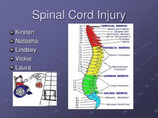
Effects of Spinal Cord Injury on Motoneuron Morphology. School of Life Sciences & Center for Adaptive Neural Systems, Ira A. Fulton School of Engineering Arizona State University Tempe, AZ. Ashley Diamond Ashley.Diamond@asu.edu. Spinal Cord Injury Background Information:.

E N D
Effects of Spinal Cord Injury on Motoneuron Morphology School of Life Sciences & Center for Adaptive Neural Systems, Ira A. Fulton School of Engineering Arizona State University Tempe, AZ Ashley Diamond Ashley.Diamond@asu.edu
Spinal Cord Injury Background Information: • Most spinal cord injuries are classified as spinal cord contusions (Spinal Cord 2007) • The motoneuron somas (cell bodies) become quite enlarged as a result of the injury. (Bose et al. 2005) • Supraspinal control is impaired in Spinal Cord injury and muscle atrophy can occur. (Barbeau et al. 2002a; Dietz 1992)
Spinal Cord Injury Continued.. • Passive exercise can regularize specific spinal reflexes in the absence of supraspinal control (Skinner et al. 1996). This would essentially prevent muscle atrophy by exciting motor pathways. • Active exercise can be induced through electrical stimulation therapy, by implanting electrodes into the motor points of muscles involved in basic flexion and extension needed for walking. • However, the effects of rehabilitative therapy on motoneuron pool densities and morphology can not be assessed without first mapping the lumbar spinal cord before and after injury.
Objective: To reconstruct spinal cord morphology from histological sections of the lumbar regions containing fluorescently labeled motoneurons of four muscles. Significance: The reconstructions will provide a map of neuron pools and quantitative data for soma sizes so that the effects of Spinal Cord Injury on motoneuron morphology and pool densities can be assessed in future experiments.
Spinal Cord Injury Model SCI is mimicked by implementing a T-8 dorsal contusion in a rodent.
Muscles were injected with a fluorescent labeled Choleratoxin beta sub unit (CTβ) and Alexfluor 594 Red/Green in order to fluoresce corresponding motoneuron pools Red: Tibialis Anterior, Illiacus Green: Gastrocnemius Medialis, Bicep Femoralis Fairchild et al, 2007
Trace Spinal Cord Sections 2.5x magnification of 40 micrometer thick section (Z-axis) Grey matter Dorsal Horn Ventral Horn Central canal White matter 1000 microns
Control Vs. Contused Spinal Cord Reconstruction Contused Normal White matter White matter Grey matter Caudal Caudal Rostral Rostral Grey Matter Ventral Dorsal Dorsal Dorsal White matter Grey Matter Rostral side looking down Rostral side looking down Ventral Ventral
Fluorescent retrogradely labeled motoneurons Biceps Femoralis Tibialis Anterior 100 Microns 100 Microns 20x magnification of 40 micrometer thick sections of spinal cord Cell body or soma Primary Dendrites Primary Dendrites Nucleus
Neuron 4 Neuron 4 Neuron 3 Neuron 3 Neuron 1 Neuron 1 Neuron 2 Neuron 2 Mapping Pool Locations Quantitative Data % location in the mediolateral axis =A*100/(A+B) % location in the dorsoventral axis =C*100/(C+D)
Location of Motoneuron Pools in Normal Rodent Lumbar Spinal Cord Bicep Femoralis neuron pool Grey Matter Caudal White Matter Ventral Dorsal Rostral
References • Barbeau H, Ladouceur M, Mirbagheri MM, and Kearney RE. The effect of locomotor training combined with functional electrical stimulation in chronic spinal cord injured subjects: walking and reflex studies. Brain Res Brain Res Rev 40: 274-291, 2002b. • Bose P, Parmer R, Reier PJ, and Thompson FJ. Morphological changes of the soleus motoneuron pool in chronic midthoracic contused rats. Neurology. 191: 13-23, 2005. • Dietz V. Human neuronal control of automatic functional movements: interaction between central programs and afferent input. Physiology Rev 72: 33-69, 1992. • Fairchild, Mallika, M., J.W. Graham, A.V. Iarkov, D. Hagner; R. Jung. Characterization of motoneuron morphology in a complete and incomplete spinal cord injury in Rodent models. Program Number 76.4, 2007. Neuroscience Meeting Planner. San Diego, CA. Society of Neuroscience, 2007, Online (November 3rd- November 7th Poster Presentation) • Skinner RD, Houle JD, Reese NB, Berry CL, and Garcia-Rill E. Effects of exercise and fetal spinal cord implants on the H-reflex in chronically spinalized adult rats. Brain Res 729: 127-131, 1996. • Spinal Cord Injury: Definition, Epidemiology, Pathophysiology. E-Medicine. 21 February 2007. http://www.emedicine.com/pmr/topic182.htm • Thota AK, Watson SC, Knapp E, Thompson B, and Jung R. Neuromechanical control of locomotion in the rat. J Neurotrauma 22: 442-465, 2005
Thank you! Ranu Jung, PhD Director, Center for Adaptive Neural Systems, ASU James Lynskey, PhD, PT Center for Adaptive Neural Systems, ASU and AT Still University Alex Iarkov, PhD Center for Adaptive Neural Systems, ASU Seung-Jae Kim, PhD Center for Adaptive Neural Systems, ASU Mallika Fairchild Center for Adaptive Neural Systems & Harrington Dept. of Bioengineering, ASU Brian Hillen Center for Adaptive Neural Systems & Harrington Dept. of Bioengineering, ASU Funding: • NASA Space Grant • NIH • Center for Adaptive Neural Systems;IRA A. Fulton School of Engineering at ASU