CELL DIVISION
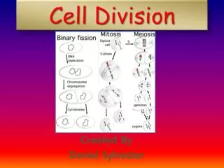
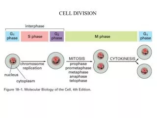
CELL DIVISION. Stages of mitosis (animal cell) prophase: - chromosomes condense (replicated in S phase) - centrosomes separate (duplicated in S phase) prometaphase: - nuclear envelope breaks down - MTs contact chromosomes, spindle forms. metaphase:

CELL DIVISION
E N D
Presentation Transcript
Stages of mitosis (animal cell) prophase: - chromosomes condense (replicated in S phase) - centrosomes separate (duplicated in S phase) prometaphase: - nuclear envelope breaks down - MTs contact chromosomes, spindle forms
metaphase: - chromosomes align at spindle equator (metaphase plate) anaphase: - sister chromatids separate - chromosomes move to poles - poles move apart telophase: - nuclear envelope reforms - chromosomes decondense - interphase array of MTs reforms cytokinesis: - contractile ring pinches cell in two
Centrosome cycle (animal cell): - centrosome (centriole) duplication begins at the start of S phase - remains as one complex until M phase In early embryonic cells, the centrosome cycle can operate without a nucleus - egg cell extracts
Mediators of mitotic chromosome structure: - cohesins: deposited along the length of sister chromatids as the DNA is replicated - hold sisters together - condensins: coil DNA - mediate chromosome condensation
Structure of a spindle: 3 classes of MTs (polar MTs) How does a spindle form, and how does it work to separate chromosomes?
Prophase: changes in MT dynamics - more MTs nucleated from centrosome - shorter, more dynamic MTs Quantifying MT dynamics: - inject fluorescent tubulin - bleach with laser - measure recovery (newly formed MTs) - t1/2 = time to 50% recovery
MT dynamics: regulated by MAPs vs. catastrophins centrosomes incubated in Xenopus egg extracts higher catastrophe rates = shorter MTs
Spindle formation in vitro: mitotic extracts + DNA + centrosomes - abnormal spindles form when ratio of MAPs:catastrophins is perturbed no MAP (MTs are too short)
Centrosome separation in prophase is driven by plus-end motors (KLPs) - balanced by minus-end motors
Yeast mutants: identification and characterization of spindle motors (-) (+)
Prometaphase: kinetochores capture MTs (mechanism of attachment??)
Forces that drive chromosomes to metaphase plate: - kinetochores pull chromosomes to poles: (-)end directed motors? - astral ejection force: (+)end directed motors on chromosome arms
Metaphase: - chromosomes continue to oscillate at metaphase plate (vertebrate cells) - MTs undergo poleward flux (function?)
- poleward flux of metaphase MTs can be measured with caged fluorescein
Dynamics of individual MTs can be measured with fluorescence speckle microscopy: - poleward flux of metaphase MTs occurs in kinetochore and overlap MTs but not in astral MTs
Anaphase A: - kinetochore MTs shorten - chromosomes move to poles Fluorescent tubulin injections show locations of MT growth, depolymerization
Anaphase B: - poles separate - overlap MTs lengthen
Bipolar spindles can assemble without centrosomes or chromosomes
What determines the position of the cleavage furrow? - signal from asters to cortex - signal from central spindle - chosen before mitosis (position of spindle from previous mitosis)
Contractile ring of actin and myosin: red = actin, green = myosin II