Download
1 / 106
1.11k likes | 1.55k Vues
Explore the distinctions between Gram-positive and Gram-negative cell walls, the classification of Gram-negative bacteria according to phenotypic similarities, and the role of various bacterial groups such as Proteobacteria in different ecosystems. Discover symbiotic relationships between plants and bacteria like Azospirillum, Rhizobium, and Agrobacterium. Uncover how bacteria like Nitrobacter aid in ammonia conversion aerobically.

E N D
Differences Between Gram Positive and Gram Negative Cell Wall The Gram positive cell wall is thick and composed of many layers of peptidoglycan that are bound together by teichoic acids and lipoteichoic acids. The Gram negative cell wall has a thinner layer of peptidoglycan. There is an outer membrane with lipopolysaccharide on the outer surface, and a periplasmic space between the inner and outer membranes.
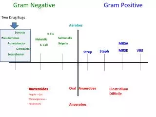
Gram Negative Bacteria (Grouped according to phenotypic similarities, as in Bergey’s Manual, 9th ed.) Group 1 The Spirochetes (Treponema, Borrelia) Group 2 Aerobic/Microaerophillic, Motile, Helical/Vibroid (Azospirillum, Bdellovibrio, Campylobacter) Group 3 Nonmotile, Curved Bacteria Group 4 Aerobic/Microaerophillic Rods and Cocci (Agrobacterium,Azotobacter, Azomonas,Bordetella, Legionella,Neisseria,Pseudomonas,Rhizobium) Group 5 Facultative Anaerobic Rods (Enterobacteriaceae, Vibrionaceae, Pasteurellaceae) Group 6 Anaerobic, Straight, Curved and Helical Bacteria (Bacteroides) Group 9 The Rickettsias and Chlamydias Group 12 Aerobic Chemolithotropic Bacteria (Nitrobacter,Nitrosomonas, Thiobacillus)
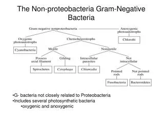
Gram Negative Bacteria Formerly called Gracilicutes, phyla have been reorganized so the bacteria are grouped according to similarities seen in their 16 rRNA sequences. The phylum = ProteobacteriaClassAlphaproteobacteria - Azospirillum - Rhizobium - Agrobacterium - Nitrobacter - Purple Non-sulfur bacteria - Acetobacter and Gluconobacter - Brucella - Rickettsia ClassBetaproteobacteria - Nitrosomonas - Neisseria - Bordetella - Burkholderia - Thiobacillus - Spirillum Spirillum
ClassDeltaproteobacteria - Desulfovibrio - Bdellovibrio - Myxobacteria ClassEpisilonproteobacteria - Campylobacter - Helicobacter Gram Negative Bacteria (grouped according to similarities seen in their 16 rRNA sequences) ClassGammaproteobacteria - Purple sulfur bacteria - Legionella - Coxiella - Methane oxidizers - Glycolytic Facultative Anaerobes Enterobacteriaceae Vibrionaceae Pasteurellaceae - Pseudomonas - Azotobacter and Azomonas Minor PhylaChlamydiae Spirochaetes - Treponema - Borrelia Bacteroidetes
Gram Negative Bacteria the Alphaproteobacteria
Bacteria that are symbiotic with plants Azospirillum, Rhizobium, Agrobacterium Azospirillum – Nitrogen fixing association with the roots of tropical grasses including sugar cane. The bacteria colonize the surface of the grass roots. Rice growth enhanced by compost containing nitrogen fixing bacteria, phosphate binding and proteolytic bacteria. (Taiwan Agricultural Chemistry Division) Rhizobium – Forms an endosymbiotic nitrogen fixing association with roots of Legumes (peas, beans, clover, alfalfa). The bacteria colonize plant cells within root nodules. The bacteria converts atmospheric nitrogen N2 to ammonia (NH3) and then provides organic nitrogenous compounds such as glutamine or ureides to the plant. The plant provides the bacteria organic compounds made by photosynthesis.
Rhizobium leguminosarum There are several different strains of Rhizobium leguminosarum thatform root nodules on various legumes. The host range of a given strain ofRhizobium bacteria is determined by nod genes that are on plasmids. The symbiotic bacteria export nitrogenous compounds, such as amino acids and ureides, to the plant while the plant provides the bacteria with carbon compounds such as succinate. Rhizobium leguminosarum bv. phaseoli forms root nodules on common beans. Rhizobium leguminosarum bv. viciae forms nodules on peas. Rhizobium leguminosarum bv. trifolii forms nodules on clover.
Sinorhizobium meliloti Sinorhizobium meliloti forms root nodules on Medicago sativa (alfalfa), a common forage legume. The symbiotic bacteria fix nitrogen. (Converts N2 to organic nitrogenous compounds such as amino acids).
Bradyrhizobium japonicum Bradyrhizobium japonicum is a slow-growing strain thatforms root nodules on soybeans. Bradyrhizobium japonicum bacteroids within a soybean root nodule cell.
Bacteria that form symbiotic relationships with Plants Azospirillum, Rhizobium, Agrobacterium Agrobacterium – Infects physical wounds of dicotyledenous plants. Plasmid DNA is transferred from the bacterium into plant cells causing the formation of tumors called crown gall. Crown gall tumor tissue is modified by bacterial DNA to produce and excrete unusual amino acids called opines that the bacterium uses for food. This ability to genetically modify plant cells has been used to produce genetically modified crops. A segment of bacterial plasmid DNA Crown Gall is transferred into a plant cell.
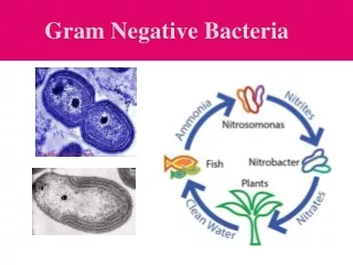
Nitrobacter Nitrifying bacteria that help to convert ammonia to nitrate aerobically. (Chemolithotrophic Metabolism) First, ammonia (NH3) is oxidized to nitrite (NO2-) by Nitrosomonas sps. Then Nitrobacter oxidizes the nitrite (NO2-) to nitrate (NO3-).They can use CO2 as a carbon source and can grow in the absence of both organic matter and sunlight. (ex: Nitrobacter winogradskyi) Purple Non-sulfur bacteria (Rhodospirillaceae) These bacteria can carry out anoxygenic photosynthesis, they can use light energy to produce a PMF. Or they can grow using organic nutrients in the dark. Rhodospirillum rubrum Picture on left shows the bacterium, note the extensive membranes. Picture on the right shows the effect of light on growth of R. rubrum. Tube on left grown in light, tube on right grown in the dark. Pigment production and anaerobic growth is seen in the light.
Gram Negative Bacteria - Alphaproteobacteria - 1 Which description is the best fit for Sinorhizobium meliloti? A. Nitrogen-fixing bacteria that form a symbiosis with beans, clover and peas. The bacteria live inside plant cells in the root nodules. B. Nitrogen-fixing bacteria that form a symbiosis with tropical grasses including sugar cane. The bacteria live on the surface of the roots. C. Slow-growing, nitrogen-fixing bacteria that form a symbiosis with soybeans. The bacteria live inside plant cells in the root nodules. D. Nitrogen-fixing bacteria that form a symbiosis with alfalfa. The bacteria live inside plant cells in the root nodules. E. Chemoautotrophic bacteria that carry out anaerobic respiration oxidizing nitrite (NO2-) to nitrate (NO3-). (Nitrobacter sps.)
Gram Negative Bacteria - Alphaproteobacteria - 1 Which description is the best fit for Sinorhizobium meliloti? A. Nitrogen-fixing bacteria that form a symbiosis with beans, clover and peas. The bacteria live inside plant cells in the root nodules. (Rhizobium leguminosarum) B. Nitrogen-fixing bacteria that form a symbiosis with tropical grasses including sugar cane. (Sweet) The bacteria live on the surface of the roots. (Azospirillum sps.) C. Slow-growing, nitrogen-fixing bacteria that form a symbiosis with soybeans. The bacteria live inside plant cells in the root nodules. (Bradyrhizobium japonicum) D. Nitrogen-fixing bacteria that form a symbiosis with alfalfa. The bacteria live inside plant cells in the root nodules. (Sinorhizobium meliloti) E. Chemoautotrophic bacteria that carry out anaerobic respiration oxidizing nitrite (NO2-) to nitrate (NO3-). (Nitrobacter sps.)
Acetobacter and Gluconobacter Used to make vinegar, incomplete oxidation of ethanol to acetic acid. This is an aerobic process and is regarded as incomplete respiration. Examples: Acetobacter acetiGluconobacter oxidans Acetobacter species, in addition to being used to make vinegar, form nitrogen-fixing symbioses with some tropical plants including: sugar cane, tea, coffee, bananas and pineapples.
Brucella Brucella abortus is an intracellular parasite of endothelial cells. It infects cattle but can spread to humans. It mostly causes a mild or asymptomatic disease in cattle but in complicated cases it can cause a cow to loose her unborn calf. Brucella is a Gram-negative pathogen that is distinguished from most other disease-causing bacteria because it does not have "obvious virulence factors" like "capsules, fimbriae, flagella, cytolysins, exotoxins, exoproteases, or other exoenzymes. Brucella is exceedingly well adapted to living within compartments in phagocytes and other cells. It is found all over the world and infects many different types of mammals including marine mammals, horses and humans. Left: Brucella cells multiplying within a cell.Image from: microwiki.kenyon.edu
Brucella abortus • Brucellosis, or Undulant Fever, is a genitourinary tract infection of sheep, cattle, pigs and other animals including humans. • B. abortus enters the body through breaks in the mucous membranes of the digestive or respiratory tracts but does not colonize the gut. It is an example of a systemic disease of the cardiovascular system that may be encountered in food or milk. B. abortus lives as an intracellular parasite in the uterus, placenta and epididymis. • The disease is usually mild but can cause spontantous abortions in pregnant mammals.
Rickettsia Very small obligate intracellular parasites that can absorb ATP from their host cells. They cannot be cultured outside host cells. They are transmitted by arthropods especially lice and ticks. R. prowazekii causes typhus.It is transmitted by lice. R. rickettsii causes RockyMountain Spotted Tick Fever. to the left:Rickettsia cells (stained red)within human epithelial cells
Epidemic Typhus Disease Symptoms of typhus include: severe headache, a sustained fever of 39°C (102 °F), cough, a rash that begins on the chest about five days after the fever appears and then spreads to the arms and legs, severe muscle pain, chills, low blood pressure, sensitivity to light, delirium and death. Ecology - A zoonotic reservoir for Rickettsia prowazekii bacteria is the Flying Squirrel. - Human to human transmission of the bacteria by lice is increased by: poverty, crowding, poor hygiene and poor nutrition, as seen in wars, refugee camps, prisons and concentration camps. History - 1546 Fracastoro described typhus in his treatise, De Contagione et Contagiosis Morbis. - In 1577 – 1579 an outbreak of Gaol Fever killed about 10% of the English population. - During Napoleon’s campaign in Russia in 1812, more of his soldiers died of typhus than were killed in battle. - In 1916 Henrique da Rocha Lima identified the bacteria that causes typhus and named it after Howard Ricketts and Stanislaus von Prowazek who had died studying epidemics. - During World War 1, delousing stations were set up on the Western front, but the disease killed about 3 million on the Eastern front.
Rocky Mountain Spotted Fever Causative Microorganism: Rickettsia rickettsii Rickettsia rickettsii is a small (~0.5 mm), Gram negative, obligate intracellular pathogen that dies quickly when outside a host cell. (ATP parasite) Transmission is by ticks of the Dermacentor family, which bite rodents and, occasionally, humans. Rocky Mountain Spotted Fever is most commonly seen in: North Carolina, Tennessee, Missouri, Arkansas, Virginia, Maryland, Oklahoma and South Carolina. It’s rarely seen in the Rocky Mountains but was first noticed in the Idaho Valley in ~1902.(This bacterium was first identified by Howard Ricketts, who died in 1910 as a result of his research, it was then described in detail by Burt Wolbach in 1919.) Symptoms: non-itchy spotted rash, fever, headache,chills, muscle aches, nausea, vomiting and petechiae.
Rocky Mountain Spotted Fever Right: Life cycle ofRickettsia rickettsii Below: left: Infected cell center: Spotted skin right: Tick From: web.uconn.edu From: en.wikipedia.org From: bio.davidson.edu From: ticksinca.blogspot.com
Gram Negative BacteriaAlphaproteobacteria - 2 Which description is the best fit for Brucella abortus? A. A tiny, obligate-intracellular parasite that is transmitted by ticks and causes Rocky Mountain spotted tick fever. B. A bacterium that can live in the cytoplasm of phagocytes and other types of cells, it infects many different types of mammals including cows and humans. The disease is usually mild but can cause fetal death in pregnant mammals. C. A tiny, obligate-intracellular parasite that is transmitted by human body lice and causes typhus. D. An aerobic bacterium that is used to make vinegar. It carries out an incomplete oxidation of ethanol to form acetic acid.
Gram Negative BacteriaAlphaproteobacteria - 2 Which description is the best fit for Brucella abortus? A. A tiny, obligate-intracellular parasite that is transmitted by ticks and causes Rocky Mountain spotted tick fever (Rickettsia rickettsia) B. A bacterium that can live in the cytoplasm of phagocytes and other types of cells, it infects many different types of mammals including cows and humans. The disease is usually mild but can cause fetal death in pregnant mammals. (Brucella abortus) C. A tiny, obligate-intracellular parasite that is transmitted by human body lice and causes typhus (Rickettsia prowazekii) D. An aerobic bacterium that is used to make vinegar. It carries out an incomplete oxidation of ethanol to form acetic acid. (Acetobacter aceti)
Gram Negative Bacteria the Betaproteobacteria
Neisseria • - bean shaped diplococci • Gram stain results may be misleading as they sometimes appear to be purple • Neisseria gonorrhea (gonococcus) is a common sexually transmitted disease • Neisseria meningitidiscauses meningitis, a severe Neisseria gonorrhea cells inflammation of the tissue that in a white blood cell (PMN). • covers the spinal cord & brain.
Gonorrhea The usual symptoms of gonorrhea in men are: burning sensation when urinating and a pus discharge from penis. Women, on the other hand, may be asymptomatic (occurs in about one half of cases) or have vaginal discharge and pelvic pain. In both men and women, if gonorrhea is left untreated, it may spread. Local spreading can cause epididymitis and sterility in males or pelvic inflammatory disease (PID) in females. In some cases the infection can spread throughout the body, affecting joints and heart valves. Gonorrhea - pus discharge
Neisseria gonorrhoeae is a Gram negative, bean-shaped, diplococcus that often gives unreliable Gram stain results. Transmission by nonsexualmeans is rare. The incubation period is 1 – 3 days. Gonorrhea usually presents as urethritis in males, causing painful urination, itching and a foul-smelling pussy exudate. More than 80% of infected women are asymptomatic but gonorrhea can cause inflammation of the cervix and uterine tubes and, if left untreated, this may progress to PID (Pelvic Inflammatory Disease) involving the uterus, ovaries and uterine tubes. (There are other causes of PID.)
Meningococcal meningitis • Neisseria meningitidis • Gram neg. diplococcus(makes endotoxin) • 1-7 day incubation period; contagious via droplets from infected person • Symptoms: aches, fever, stiff neck, petechiae, rash • Treated with Ceftriaxone & chloramphenicol • A vaccine is available
Bordetella B. pertussis causes whooping cough, a respiratory infection that can be very serious in young children. (The “P” in the DPT vaccination stands for Pertussis.)
Pertussis (Whooping Cough) - Caused by Bordetella pertussis, a small, aerobic, nonmotile, Gram negative, coccobacillus. Significant virulence factors include: - Filamentous hemagglutinin (an adhesin) - Pertussis toxin (both adhesion and toxin, it causes increased mucous production) - Adenylate cyclase toxin (increases mucous production, inhibits leukocyte movement, phagocytosis and the killing of bacteria) - Dermonecrotic toxin (causes localized hemorrhage and cell death) - Trachael cytotoxin (slows cilia movement) - Pertussis is highly contagious, spread through the air by microdroplets that are caused by coughing. It can be fatal in small children. Antibacterial drugs have little effect on the course of the disease. Vaccination uses an inactivated toxin.
Whooping Cough – Pertussis Bordetella pertussisis an aerobic Gram negative rod-shaped bacterium. The infection is transmitted by airborne aerosol. The bacteria attach to ciliated cells and colonize the trachea, bronchi, bronchioles. Disease tends to be mild in older children and adults. It begins with a runny nose and progresses to violent coughing, often with vomiting. Airways become constricted by mucus, coughing occurs 2-3 times without inhaling, followed by a struggle to inhale which produces the whooping sound. Patients become anoxic if bronchioles are obstructed. Reduced O2 in the blood can cause an increased blood pressure which can lead to hemorrhages in the eyes and brain. Since 1996 the vaccine has been based on an acellular component. The shot is now called DTaP and is regarded as less likely to cause adverse reactions than the original attenuated vaccine (DPT). Treatment with erythromycin may help if it is given very early. Most treatment is supportive because recovery depends more on regeneration of tracheal epithelium, not on the number of bacteria.
Nitrosomonas Nitrifying bacteria that converts ammonia to nitrite aerobically. This is part of nitrification. Nitrition (production of nitrite) 2NH3 + 3O2 2NO2- + 2H+ + 2H2O Example: Nitrosomonas europaea
Methane oxidizers (methanotrophs) Methane oxidizing bacteria live in the aerobic layer of pond sediment that is just above the anaerobic sediments where methanogens live. Methanogens are Archaea that make methane under anaerobic conditions. Methanotrophs can use methane as a food source. Example:Methylobacillus flagellatus(classified in beta-proteobacteria) Winogradski columns
Gram Negative Bacteria Betaproteobacteria Which description is the best fit for Bordetella pertussis? A. causes whooping cough B. causes inflammation of the central nervous system C. can oxidize ammonia to nitrite (NO2-) D. causes an STD that is characterized by a pussy exudate from the urethra
Gram Negative Bacteria Betaproteobacteria Which description is the best fit for Bordetella pertussis? A. causes whooping cough (Bordetella pertussis) B. causes inflammation of the central nervous system (Neisseria meningitidis) C. can oxidize ammonia to nitrite (NO2-) (Nitrosomonas sps.) D. causes an STD that is characterized by a pussy exudate from the urethra (Neisseria gonorrhoeae)
Gram Negative Bacteria the Gammaproteobacteria
Gammaproteobacteria Azotobacter and Azomonas These bacteria form nitrogen-fixing symbiosis with plant roots. Azotobacter vinelandia forms an association with Ash trees.
Pseudomonas - respiratory metabolism, they don’t ferment glucose anaerobically - many species are involved with rotting plant material - Pseudomonas syringae involved in nucleating ice crystals - Pseudomonas aeruginosa involved in wound and burn infections, produces a blue-green pigment that can lead to blue-pus. P. aeruginosais also frequently involved in “swimmer’s ear”. Pseudomonas aeruginosa P. aeruginosa makes a P. aeruginosa burn infection flagella stain blue-green soluble pigment.
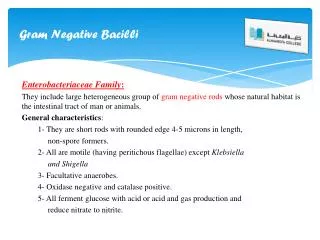
Glycolytic Facultative Anaerobes Enterobacteriaceae - entero: intestines Many Enterobacteriaeceae are found in the mammalian large intestine. - facultative anaerobes that can ferment glucose - oxidase negative, catalase positive examples include: Escherichia coli, Salmonella typhi, Enterobacter aerogenes, Klebsiella pneumonia,Shigella dysenteriae, Yersinia pestis, Serratia marcescens
Traveler’s Diarrhea E. coli common cause Coliforms Lactose fermenters Transmitted via fecal/oral route Rehydrate patient
Pathogenic Strains of Escherichia coli Virulent strains have genes for fimbriae, adhesins and a variety of toxins. O157 : H7 produces a Shiga-like toxin. This toxin can kill cells, cause kidney failure and death. E. coli and related bacteria constitute about 0.1% of gut flora. E. coli cells are able to survive outside the body for a limited amount of time, which makes them ideal indicator organisms to test environmental samples for fecal contamination.
Escherichia coli 0157
Salmonella typhi Salmonella bacteria live in bird, reptile and mammalian intestines.Salmonella bind to intestinal cells, inject proteins into the host cell that cause a normally nonphagocytic cell to engulf the bacteria. The bacteria grow inside endocytic vesicles, eventually killing the host cell. Some strains enter the blood and spread throughout the body. Salmonella Invasion
Salmonellosis Salmonella species- especially S. typhi Transmitted in contaminated food or water (fecal-oral) Causes diarrhea Typhoid fever is aninfection of the blood Incubation: 8 – 48 hrs. Typhoid symptoms:fever, headache, muscle pain,malaise, loss of appetite,rose spot rash, intestinal hemorrhage, kidney failure,peritonitis
Shigellosis Several Shigella species Bacillary dysentery(bloody diarrhea) Associated with poor sanitation Not killed by stomach acid Shiga toxin Treatment includes rehydrate patient
Shigellosis Shigella bacteria invade cells of the intestinal epithelium.Shiga toxin disrupts host cell protein synthesis. Mortality rates can be as high as 20%.