Download
1 / 53
530 likes | 593 Vues
Explore the pathways and tracts of the nervous system, including sensory and motor pathways, neuron functions, ganglia types, and conditions like Shingles. Learn about direct and indirect motor pathways for precise movements.

E N D
Pathway • route followed by a nerve impulse as it travels through the nervous system • a reflex arc is simplestof these pathways
Pathways • Include: - nerve fibers in nerves; bundles called fascicles - nerve fibers inside the brain and spinal cord; bundles called tracts - bundles of nerve fibers linking the two halves of the brain called commissures
Pathways (cont.) • sensory - ascending • motor - descending
Sensory Pathways(ascending) • start at sensory receptors and end in cerebral cortex of brain • Consist of a series of three neurons: - first-order neurons - second-order neurons - third-order neurons
First-Order Neurons • sensory neurons that convey impulses from sensory receptors to CNS • extend up to medulla on same side of body • axon terminals form synapses with second-order neurons
Second-Order Neurons (associated neurons) • carry sensory impulses to the thalamus (integrating center) • axon of the second-order neuron crosses to the opposite side of the medulla to thalamus • in thalamus, axon terminals of second-order neurons synapse with third-order neurons
Third-Order Neurons (associated neurons) • carry impulses from thalamus to cerebral cortex (where conscious sensation is produced)
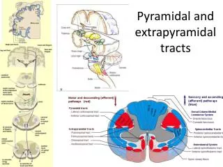
Motor Pathways(descending) • start in brain and terminate at muscles or glands • consist of upper and lower motor neurons • Two basic pathways: - direct or pyramidal -indirect or extrapyramidal
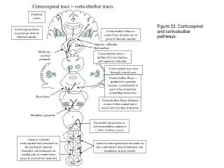
Direct Motor Pathways(pyramidal) • carry impulses from cerebral cortex directly to lower motor neurons • simplest pathway consists of two neurons; upper and lower motor neurons
Upper Motor neurons (pyramidal) • in cortex • fibers pass through bulges called pyramids on medulla oblongata - hence the name • conduct impulses from motor cortex to motor nuclei of the cerebral nerves or to the ventral gray columns of spinal cord
Lower Motor Neurons (pyramidal) • peripheral neurons whose cell bodies lie in the ventral gray column of spinal cord and terminate in skeletal muscles • responses are semivoluntary and automatic
Direct Pathway Impulses • Channeled into three tracts: - lateral corticospinal tracts - anterior corticospinal tracts - corticobulbar tracts direct pathways result in precise voluntary movements
Indirect Motor Pathways(extrapyramidal) • impulses follow complex polysynaptic circuits • carry lower motor neurons through other parts of brain • Pathways involve: - motor cortex, basal ganglia, thalamus, cerebellum, reticular formation, nuclei in the brain stem
Tracts • bundles of nerve fibers (axons) in CNS • Types of tracts: -sensory or ascending contain nerve fibers that carry impulses up the spinal cord to the brain - motor or descending contain nerve fibers that carry impulses down SC
Tracts (cont.) • Tracts are named according to: - location in spinal cord - origin - termination
Ganglia (ganglion - singular) • group of neuron cell bodies • located outside central nervous system in the peripheral nervous system
Four Basic Types of Ganglia • posterior root ganglia • sympathetic trunk ganglia • prevertebral ganglia • terminal ganglia
Posterior Root Ganglia(dorsal root ganglia) • contain cell bodies of sensory nerves • located near spinal cord in posterior (dorsal) roots of spinal nerves
Sympathetic Trunk Ganglia (sympathetic division) • form a chain of ganglia on each side of the vertebral column • extend from neck to coccyx • contain cell bodies of postganglionic sympathetic neurons • also known as paravertebral ganglia and sympathetic chain ganglia • can be dissected out separately like a string of pearls
Prevertebral Ganglia(sympathetic division) • located anterior to vertebral column • close to the abdominal arteries • contain cell bodies of postganglionic sympathetic neurons • also called collateral ganglia
Terminal Ganglia(parasympathetic division) • located near or inside internal organs (visceral effectors) • consist of clusters of cell bodies of postganglionic parasympathetic neurons • also known as intramural ganglia
Shingles or Herpes Zoster • caused by same virus that causes chicken pox (Herpes varicella-zoster) • chicken pox virus may survive in dormant state in dorsal root ganglia • stress or advancing age may cause the virus to become active
Shingles or Herpes Zoster (cont.) • virus is present in sensory trunk but damage caused by the virus is seen in the skin over the affected nerve • Symptoms: - painful raised red lesions - follow course of nerve on skin external to it - no specific treatment
Shingles or Herpes Zoster (cont.) • if 7th. cranial nerve is affected, Bell’s palsy (facial paralysis) results • if optic nerve is affected blindness will occur
Spinal Nerve • Attached to spinal cord by: - dorsal (posterior) root is composed of sensory fibers - ventral (anterior) root is composed of motor fibers
Dorsal Root (posterior) • exhibits small enlargement called dorsal root ganglionwhichcontains cell bodies of sensory neurons
Dorsal and Ventral Roots • pass laterally from spinal cord • merge to form single mixed spinal nerve • pass through intervetebral foramen (IVF) • after passing IVF spinal nerve divides into two main branches - large ventral (anterior) ramus - smaller dorsal (posterior) ramus
Spinal Cord Showing Roots and Ganglia spinal cord posterior root ganglion (dorsal) sympathetic trunk ganglion
Spinal Cord Showing Roots and Ganglia spinal cord posterior root ganglion posterior root spinal nerve sympathetic trunk ganglion vertebrae anterior root
Nuclei • located in brain or spinal cord • cluster of neuron cell bodies • CNS nuclei are isolated regions of gray matter • located within white matter of brain and spinal cord • neurons in a given nucleus perform specific functions
Basal Ganglia (cerebral nuclei or basal nuclei) • several groups of nuclei • located within white matter of cerebral hemispheres • they integrate semi-voluntary automatic movements like walking, swimming, and laughing
Thalamus • consists of a pair of oval masses on each side of 3rd ventricle in diencephalon • mostly gray matter • made up of many nuclei • Functions include: - language, memory, emotion, integration and relay of sensory impulses to the cerebral cortex
Ventricles • four cavities within brain - two lateral ventricles - third ventricle - fourth ventricle • each ventricle contains capillary network called choroid plexus which forms cerebrospinal fluid from blood plasma
Hypothalamus • in region of diencephalon • located below two halves of thalamus • consists of a variety of nuclei and nuclear areas - most important control area for internal environment • Functions: - thirst, hunger, hormone production, and fear and rage reactions
Brain Stem • nuclei for most of the cranial nerves are located in brain stem • other nuclei located in brain stem controlbreathing, the force and rate of heart contractions, and blood vessel diameter
Cerebellum • cerebellar nuclei are regions of gray matter located deep within cerebellum • Concerned with: - balance, proprioception, (self-awareness), and the planning and coordination of complex muscular activities
Nuclei 3rd. Ventricle 2 divisions of lenticular nucleus head of caudate nucleus posterior lateral nucleus of thalamus tail of caudate nucleus medial pulvinar nucleus of thalamus choroid plexus
Reflex (stimulus) • fast, predictable, automatic, unconscious response to change in the environment that helps to maintain homeostasis • occurs in gray matter • Change can be: - external - outside the body - internal - inside the body
Homeostasis • internal environment of body is maintained at a relatively constant level • blood pressure, plasma glucose, pH, and body temperature are examples of body conditions that must be consistently maintained
Homeostatic Mechanism • sequence of events that maintains a consistent internal environment • homeostatic mechanisms are called negative feedback control systems
Negative Feedback • principle dictating most control systems • response in which a stimulus counteracts, reverses or reduces original stimulus (back to original value)
Types of Reflexes • somatic reflexes: - involve contraction of of skeletal muscles • autonomic reflexes: - involve the contraction of smooth muscles, cardiac muscles, and glands
Reflex Arc • basic structural and functional unit of nervous system • begins with a receptor at end of a sensory nerve fiber
Sequence of Events in Response to Stimulus • receptor • sensory pathway • integrating center • motor pathway • effector
Receptor • specialized sensory nerve ending • detects environmental change (stimulus) • responds by initiating a nerve impulse in a sensory neuron
Sensory Pathway(afferent) • carries nerve impulse from receptor to central nervous system