Download

1 / 19
190 likes | 279 Vues
Explore normal and abnormal anatomy through MRI, emphasizing clinical interpretation based on patient history. Learn to read MRI reports and manipulate parameters for T1W, T2W, and PDW imaging techniques. Discover insights into spinal and musculoskeletal imaging.

E N D
Fundamentals of Image Interpretation Joseph Castillo B.Sc, M.Sc (MRI)
Five levels • Normal anatomy as seen on MR • Abnormal anatomy • Accurate and complete patient history – In our unit this is not an option. • Pathology – Read MRI reports • Accurate clinical interpretation
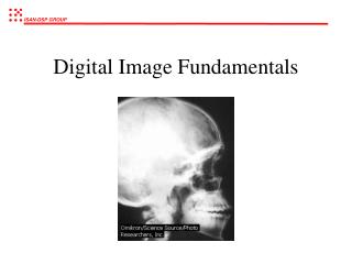
Clinical Imaging • Three basic orthogonal planes used in MRI – axial, sagittal and coronal • Unlike CT, radiographers have the ability to image biological functions not just electron density. • We manipulate parameters to get T1W, T2W and PDW. The appearance of the organ is changed.
Spine • Which one is T2w? A B