Lesson Plan: Joints 1
Lesson Plan: Joints 1. 5 minutes: Breath of Arrival and Attendance 10 minutes: Tibialis anterior and peroneus longus 40 minutes: Joints 1. Classroom Rules. Punctuality- everybody's time is precious: Be ready to learn by 9:00, we'll have you out of here by 1:30

Lesson Plan: Joints 1
E N D
Presentation Transcript
Lesson Plan: Joints 1 5 minutes: Breath of Arrival and Attendance 10 minutes: Tibialis anterior and peroneus longus 40 minutes: Joints 1
Classroom Rules Punctuality- everybody's time is precious: • Be ready to learn by 9:00, we'll have you out of here by 1:30 • Tardiness: arriving late, late return after breaks, leaving early The following are not allowed: • Bare feet • Side talking • Lying down • Inappropriate clothing • Food or drink except water • Phones in classrooms, clinic or bathrooms You will receive one verbal warning, then you'll have to leave the room.
Tibialis Anterior Origin: Upper 2/3 of lateral tibia Interosseousmembrane Insertion: Base of 1st metatarsal Medial cuneiform (plantar) Actions: Ankle dorsiflexion Foot inversion
Tibialis Anterior Origin: Upper 2/3 of lateral tibia Interosseousmembrane Insertion: Base of 1st metatarsal Medial cuneiform (plantar) Actions: Ankle dorsiflexion Foot inversion
Tibialis Anterior Origin: Upper 2/3 of lateral tibia Interosseousmembrane Insertion: Base of 1st metatarsal Medial cuneiform (plantar) Actions: Ankle dorsiflexion Foot inversion
Tibialis Anterior Origin: Upper 2/3 of lateral tibia Interosseousmembrane Insertion: Base of 1st metatarsal Medial cuneiform (plantar) Actions: Ankle dorsiflexion Foot inversion
Peroneus Longus Origin: Proximal 2/3 of lateral fibula Insertion: Base of 1st metatarsal Medial cuneiform (plantar) Actions: Ankle plantarflexion Foot eversion
Peroneus Longus Origin: Proximal 2/3 of lateral fibula Insertion: Base of 1st metatarsal Medial cuneiform (plantar) Actions: Ankle plantarflexion Foot eversion
Peroneus Longus Origin: Proximal 2/3 of lateral fibula Insertion: Base of 1st metatarsal Medial cuneiform (plantar) Actions: Ankle plantarflexion Foot eversion
Peroneus Longus Origin: Proximal 2/3 of lateral fibula Insertion: Base of 1st metatarsal Medial cuneiform (plantar) Actions: Ankle plantarflexion Foot eversion
Joints 1 “Opportunity is missed by most people because it is dressed in overalls and looks like work.” –Thomas Edison
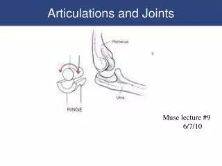
Joints Joint (AKA: articulation or arthrosis)Where bones come together or join.
Joints Physiology • Enable the body to move. • Bear the weight of the body. • Provide stability.
Structural and Functional Classification Fibrous / Synarthrotic Cartilaginous / Amphiarthrotic Synovial / Diarthrotic
Structural and Functional Classification Fibrous / Synarthrotic • Connected by dense fibrous connective tissue, consisting mainly of collagen. • Extremely limited movement. • Examples: cranial sutures, facial sutures, teeth, and tibiofibularjoints
Structural and Functional Classification Cartilaginous / Amphiarthrotic • Connected by cartilage . • Slightly movable . • Examples: costochondral joints, pubic symphysis, and intervertebral disk joints
Structural and Functional Classification Synovial / Diathrotic • Contains a joint capsule that contains synovial fluid to nourish and lubricate the articulating surfaces. • Freely movable. • Examples: glenohumeral, iliofemoral . . . see Synovial Joints section for more examples
Synovial Joints Articular cartilage Joint capsule Joint cavity Synovial membrane Synovial fluid Synovial sheath Bursa Meniscus
Synovial Joints Articular cartilage Hyaline cartilage covering an epiphysis.
Synovial Joints Joint capsule Double-layered structure around a synovial joint. The outer layer is fibrous and forms ligaments, and the inner layer is the synovial membrane.
Synovial Joints Joint cavity Space within a joint capsule. Lined with a synovial membrane.
Synovial Joints Synovial membrane Membrane that lines cavities of freely moving joints, synovial sheaths, and bursae.
Synovial Joints Synovial fluid Viscous fluid secreted by synovial membranes. Provides nutrition and lubrication.
Synovial Joints Synovial sheath Tube-like structure lined with synovial membrane that surrounds long tendons.
Synovial Joints Bursa Collapsed sac-like structure with an interior lining of synovial , membrane. Contains synovial fluid. Plural is bursae.
Synovial Joints Meniscus Fibrocartilage pads found in select joints such as the knee and jaw. Helps the joint move smoothly and serves as a shock absorber. Plural is menisci.
Types of Synovial Joints Hinge Pivot Ellipsoidal / condyloid Saddle Ball and socket Gliding / planar
Types of Synovial Joints Hinge Limited to flexion and extension .
Types of Synovial Joints Pivot Limited to rotation .
Types of Synovial Joints Ellipsoidal / condyloid Limited to flexion, extension, abduction, and adduction.
Types of Synovial Joints Saddle Allowing flexion, extension, abduction, adduction, opposition, reposition, and circumduction, but not rotation.
Types of Synovial Joints Ball and socket Allowing all movements except gliding . Offers the greatest range of motion.
Types of Synovial Joints Gliding / planar Limited to planar movements but movement may be permitted in all planes .
Types of Synovial Joints Extension Straightening or increasing the angle of a joint . Flexion Bending or decreasing the angle of a joint.
Types of Synovial Joints Flexion Bending or decreasing the angle of a joint. Extension Straightening or increasing the angle of a joint .
Types of Synovial Joints Flexion Bending or decreasing the angle of a joint. Extension Straightening or increasing the angle of a joint .
Types of Synovial Joints Hyperextension A continuation of extension beyond anatomic position.
Types of Synovial Joints Adduction Movement toward , the median plane. Abduction Movement away , from the median plane.
Types of Synovial Joints Adduction Movement toward , the median plane. Abduction Movement away , from the median plane.
Types of Synovial Joints • Pronation Medial (inward) rotation of the forearm so that the palm is turned down . Supination Lateral (outward) rotation of the forearm so that the palm is turned up .
Types of Synovial Joints Dorsiflexion Flexing the ankle dorsally so that the toes are moving toward the shin . Plantarflexion Extension of the ankle such that the toes are pointing downward , increasing the ankle angle anteriorly.
Types of Synovial Joints Eversion Elevation of the lateral, edge of the foot so that the sole is turned outward (or laterally). Inversion Elevation of the medial , edge of the foot so that the sole is turned inward (or medially).
Types of Synovial Joints Circumduction Cone -shaped range of motion that occurs when the distal end moves in a circle and the proximal end is fixed.
Types of Synovial Joints Rotation Circular movement when a bone moves around its own central axis .
Types of Synovial Joints Right and left rotation Rotationfor joints that lie within the median axis.
Types of Synovial Joints Lateral and medial rotationRotation for joints that lie outside of the median axis.
Types of Synovial Joints Upward and downward rotationRotation of the scapula so that the glenoidfossa faces either upward or downward.
Types of Synovial Joints Depression Lowering or dropping a body part. Moving inferiorly . Elevation Raising or lifting a body part. Moving superiorly .