Understanding the Skull and Skeleton: An In-depth Overview
Explore the intricate details of the human skull and skeleton, from the division of the cranium to the structure of facial bones. Learn about crucial openings, axial and appendicular skeleton, and significant bones like the sternum, ulna, and radius.

Understanding the Skull and Skeleton: An In-depth Overview
E N D
Presentation Transcript
Overview of Skull Geography • With the lower jaw removed, the skull resembles a lopsided, hollow, bony sphere. The facial bones form its anterior aspect , and the cranium forms the rest of the skull. The cranium can be divided into a vault and a base. The cranial vault, also called the calvaria, forms the superior, lateral, and posterior aspects of the skull, as well as the forehead. • The skull has about 85 named openings (foramina, canals, fissures, etc.) the most important of these provide passageways for the spinal cord, the major blood vessels serving the brain, and the 12 pairs of cranial nerves ( #’s 1-12), which transmit impulses to and from the brain.
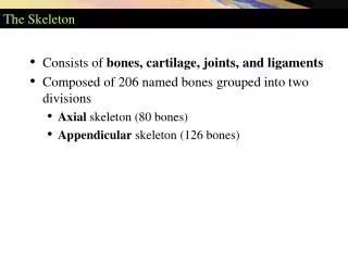
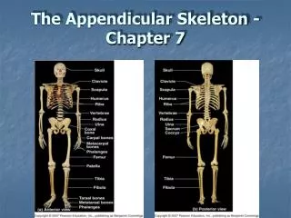
Appendicular Skeleton Axial Skeleton
The Axial Skeleton • Structured from 80 bones segregated into 3 major regions: • Skull • Vertebral Columns • Bony Thorax • This part of the skeleton supports the head, neck, and trunk, and it protects the brain, spinal cord, and the organs in the thorax
The Skull • The skull is the body’s most complex bony structure. It’s formed by cranial and facial bones, 22 in all. • Most skull bones are flat bones. Except for the mandible (jaw) which is connected to the rest of the skull by a freely movable joint
Inside the Cranium • Frontal Bone • Shell-shaped frontal bone forms the anterior cranium. It articulates posteriorly w/ the paired parietal bones via the prominent coronal suture. • The most anterior part of the frontal bone is the vertical frontal squama, commonly called the forehead.
Inside the Cranium contd….. • Parietal Bones and the Major Sutures • The two large parietal bones are curved, rectangular bones that form most of the superior and lateral aspects of the skull; hence they form the bulk of the cranial vault.
The Facial Skeleton Cameron Braddy
Facial Bones • The facial skeleton is made up of 14 different bones. Men’s faces are more elongated than women, meaning women’s faces tend to be less angular.
Vomer • Plow shaped vomer, lies in the nasal cavity. • Forms part of the nasal septum. • Discussed below in connection with the nasal cavity.
Inferior Nasal Conchae • Inferior nasal conchae are two paired bones that are thin, curved and in the nasal cavity. • Project medially from the lateral walls of the nasal cavity. • Largest of the three pairs of conchae.
The Coccyx • Commonly referred to as our tailbone. • Triangular bone • Consists of four vertebrae fused together. • Affords the pelvic organs. • Nearly useless. • Often snipped off by a physician.
Bony Thorax • Bony underpinnings of the thorax. • Roughly cone shaped. • Forms a protective cage around the vital organs. • Supports shoulders, and upper limbs. • Provides attachment points for many muscles of the neck, back, chest, and shoulders.
The Upper Limb, Arm • Thirty separate bones form each upper limb. • The humerus is the sole bone of the arm. • At the proximal end of each humerus is smooth hemispherical head, and at the distal end are two chondyles are medial trochlea.
The Sternum • The Sternum is a flat bone located in the middle of the bony thorax. • Means breastbone. • The fusion of three bones, the manubrium, the body, and the xiphoid. • Manubrium at the top, the body in the middle and the xiphoid at the bottom.
Anatomy and Physiology Chapter 7 By: Caroline Baker
Special Characteristics of orbits and nasal cavity: the orbits, naval cavity, paranasal sinuses, and hyoid bone. Page: 213- 216
The Orbits • The orbits are bony cavities in which the eyes are firmly encased and cushioned by fatty tissue • The orbits are formed by seven bones – frontal, sphemoid, zygomatic, maxilla, palatine, lacrimal, and ethmoid bones.
Nasal Cavity • The nasal cavity is constructed of bone and hyaline cartilage. The roof of the nasal cavity is formed by the cribriform plate of the ethmoid.
Paranasal Sinuses • You can see this sinuses in an x-ray image • Paranasal sinuses cluster around the nasal cavity
Hyoid Bone • U / horseshoe shaped • The hyoid bone lies just inferior to the mandible in the anterior neck. • Neck muscles that raise and lower the larynx durning swallowing and speech.
Sternum, Homeostatic imbalance Page: 226
Sternum • The sternum (breastbone) lies in the anterior midline of the thorax. • The sternum is a flat bone and it is approximately 15 cm( 6 inches) long.
Homeostatic imbalance • An inability to maintain homeostasis may lead to death or a disease, a condition known as homeostatic imbalance.( Wikipedia) • Of the sternum- in some people the xiphoid process projects dorsally
Forearm, Ulna, Radius Page: 233
Forearm (Ulna and Radius) • Two parallel long bones, the radius and the ulna. • The ulna is slightly longer than the radius. It has the main responsibility for forming the elbow joint with the humerus. • The radius (rod) is thin at its proximal end and wide distally- opposite of the ulna. • The head of the radius is shaped somewhat like the head of a nail.
Leg, Tibia , Fibula Page: 243
Leg (tibia and Fibula) • Two parallel bones, the tiba and fibula, form the skeleton of the leg, the region of the lower limb between the knee and the ankle. • The tibia (shinbone) receives the weight of the body from the femur and transmits it to the foot. • The fibula (pin) is a sticklike bone with slighty expanded ends.
Facial Bones By: Kourtnie Moore
Mandible The U- shaped or lower jaw bone. It is one of the largest, strongest bone of the face. It has a body, which forms the chin, and two upright “rami” branches.
Maxillary Bones The Maxillary Bone is also called sometimes Maxillae. They form the upper jaw and the central portion of the facial skeleton. The Maxillae carry up the upper teeth in the Alveolar Margins. The Maxillae meet medially, forming the pointed Anterior Nasal Spine at their junction. The Palatine Processes of the maxillae project posterior form the alveolar margins and fuse medially. The Frontal Processes extend superiorly to the frontal bone, forming part of the lateral aspects of the bridge of the nose. The regions the flank the nasal cavity laterally contain the Maxillary Sinuses. Laterally, the maxillae articulate with the zygomatic bones via their Zygomatic Processes. The Inferior Orbital Fissure is located deep within the orbit at the junction of the maxilla.
Zygomatic The irregularly shaped bones. Are commonly called the “cheek bones”. They join in with the Temporal Posteriorly and with the Zygomatic processes of maxilla Anteriorly. The Zygomatic Bones form the prominences of the cheeks and part of the inferolateral margins of the orbits.
PG. 220 By: Brandon Jenkins
Ligaments In order for the bone structures to stand up they have to have a system of cable like supports. The strap like ligaments and trunk muscles assume that role. There are two major supporting ligaments they are the anterior and posterior longitudinal ligaments which run a continuous band down the front and back of the spine as shown to the right.
Intervertebral Discs The intervertebral disc is a cushion like pad between each disc. It has two parts the nucleus pulpous which acts like a rubber ball and gives the disc its elasticity and the annulus fibrosus which limits the expansion of the nucleus pulpous. They act as shock absorbers when your living your everyday lives. These disc are about 25% of your spinal weight.
Homeostatic Imbalance A homeostatic imbalanceis a sudden physical trauma to the spine- for example there is a herniated disc also referred to as a slipped disc. The disc can slip and pinch a nerve causing numbness and severe pain. They can be treated with medicine but if it fails they have to do surgery.
Regional Vertebral Characteristics&General Structure • Structure • All vertebrae have a common structural pattern. • Each vertebrae consists of a body, or centrum, anteriorly and a vertebral arch posteriorly. • The vertebral arch is a composite structure formed by two pedicles (little foot) and two laminae.
Vertebral Characteristics • Flexion and extension (anterior bending and posterior straightening of the spine) • Lateral flexion (bending the upper body to the right or left). • Rotation (in which vertebrae rotate on one another in the longitudinal axis of the spine).
Stevie Peele Anatomy and Physiology
Sphenoid Bone • Butterfly-shaped • Considered the keystone of the cranium because it form the central wedge that articulates with all other cranial bones.
Sphenoid Bone Cont. • Within the body ofthe sphenoid are the paired sphenoid sinuses. • Thesuperior of the body bears a saddle-shaped prominence is the sella turcica meaning “Turk’s saddle”. • The seat of the saddle is called the hypophyseal.
Surfaces • The lateral surfacesof the body are united with the great wings and the medial pterygoid plates. Above the attachment of each great wing is a broad groove, curved something like the italic letter f; it lodges the internal carotid artery and the cavernous sinus, and is named the carotid groove. Along the posterior part of the lateral margin of this groove, in the angle between the body and great wing, is a ridge of bone, called thelingula. • The posterior surface, quadrilateral in form is joined, during infancy and adolescence, to the basilar part of the occipital bone by a plate of cartilage. Between the eighteenth and twenty-fifth years this becomes ossified, ossification commencing above and extending downward.
Sphenoid Cont. • Piercing the lesser wings of the sphenoid, the optic canals allow passage of the optic nerves from the back of each eye to enter the brain and cross at the optic chiasma above to the pituitary gland. A cleft between the greater and lesser wings of the sphenoid, the superior orbital fissure transmits several critical structures that pass between the orbit and the brain.
EthMoid Bone • The ethmoid boneis exceedingly light and spongy, and cubical in shape; it is situated at the anterior part of the base of the cranium, between the two orbits, at the roof of the nose, and contributes to each of these cavities. It consists of four parts: a horizontal or cribriform plate, forming part of the base of the cranium; a perpendicular plate, constituting part of the nasal septum; and two lateral masses or labyrinths.
Sutural Bones • Also called Wormian bones • Are tiny irregularly shaped bones or bone clusters that appear with sutures • Structurally unimportant • Numbers varies, and not all skulls exhibits them • They represent additional ossification centers that appeared when the skull was expanding very rapidly during fetal development.
Ischium Stevie Peele
Ischium Forms the posterior inferior part of the hip bone. Roughly L or arched-shaped Inferior ramus that joins pubis anteriorally Its ischial spine projects medially into the pelvic cavity and serves as a point of attachment
Ischium Just inferior to the ischeial spine is the lesser sciatic notch. A number of nerves and blood vessels pass through this notch to supply the anogenital area. The inferior surface of the ischial body is rough and grossly thickened as the ischial tuberosity.