Download
1 / 48
480 likes | 527 Vues
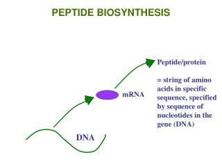
ie. amino acid peptide polypeptide protein. larger. smaller. + H 2 O. +. + H 2 O. +. Dehydration synthesis/condensation reaction. Dehydration Synthesis. Hydrolysis. Hydrolysis reaction. Carbohydrates. Differ in position & orientation of glycosidic linkage.

E N D
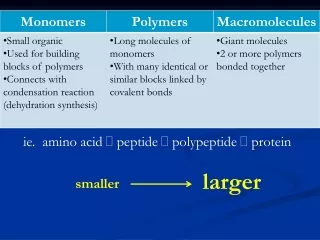
ie. amino acid peptide polypeptide protein larger smaller
+ H2O + + H2O +
Carbohydrates Differ in position & orientation of glycosidic linkage • Fueland building material • Include simple sugars (fructose) and polymers (starch) • Ratio of 1 carbon: 2 hydrogen: 1 oxygen or CH2O • monosaccharide disaccharide polysaccharide • Monosaccharides = monomers (eg. glucose, ribose) • Polysaccharides: • Storage (plants-starch, animals-glycogen) • Structure (plant-cellulose, arthropod-chitin)
. • Carbohydrates are made of carbon, hydrogen, and oxygen. • Carbohydrates include sugars and starches. • Monosaccharides are simple sugars. • Polysaccharides include starches, cellulose, and glycogen.
Polymer (starch) Starch is a polymer of glucose monomers that often has a branched structure. Polymer (cellulose) Cellulose is a polymer of glucose monomers that has a straight, rigid structure monomer • Carbohydrates can be broken down to provide energy for cells. • Some carbohydrates are part of cell structure.
The polysaccharide cellulose is a major component of the tough wall of plant cells Like starch, cellulose is a polymer of glucose, but the glycosidic linkages differ Cellulose in human food passes through the digestive tract as insoluble fiber Some microbes use enzymes to digest cellulose Many herbivores, from cows to termites, have symbiotic relationships with these microbes
Glycogen is a storage polysaccharide in animals Humans and other vertebrates store glycogen mainly in liver and muscle cells
Chitin, another structural polysaccharide, is found in the exoskeleton of arthropods Chitin also provides structural support for the cell walls of many fungi
The structureof the chitinmonomer Figure 5.9 Chitin forms the exoskeletonof arthropods. Chitin is used to make a strong and flexiblesurgical thread that decomposes after thewound or incision heals.
Storage polysaccharides of plants (starch) and animals (glycogen)
I. Proteins Myoglobin protein • “Proteios” = first or primary • 50% dry weight of cells • Contains: C, H, O, N, S
Protein Functions (+ examples)? • Enzymes (lactase) • Defense (antibodies) • Storage (milk protein = casein) • Transport (hemoglobin) • Hormones (insulin) • Receptors • Movement (motor proteins) • Structure (keratin)
Four Levels of Protein Structure • Primary • Amino acid (AA) sequence • 20 different AA’s • peptide bonds link AA’s
Amino Acid • R group = side chains • Properties: • hydrophobic • hydrophilic • ionic (acids & bases) • “amino” : -NH2 • “acid” : -COOH
Four Levels of Protein Structure (continued) • Secondary • Gains 3-D shape (folds, coils) by H-bonding • Alpha (α) helix, Beta (β) pleated sheet
Basic Principles of Protein Folding Hydrophobic AA buried in interior of protein (hydrophobic interactions) Hydrophilic AA exposed on surface of protein (hydrogen bonds)
Four Levels of Protein Structure (continued) • Tertiary • Bonding between side chains (R groups) of amino acids • H bonds, ionic bonds, disulfide bridges, hydrophobic interactions, van der Waals interactions
Four Levels of Protein Structure (continued) • Quaternary • 2+ polypeptides bond together
amino acids polypeptides protein • Bonding (ionic & H) can create asymmetrical attractions
Chaperoninsassist in proper folding of proteinsNewly made proteins usually must fold from a linear chain of amino acids into a three-dimensional form.Chaperonins belong to a large class of molecules that assist protein folding, called molecular chaperones. The energy to fold proteins is supplied by adenosine triphosphate (ATP).
Protein structure and function are sensitive to chemical and physical conditions • Unfolds or denatures if pH and temperature are not optimal
II. Nucleic Acids Function: store hereditary info
Nucleotides: monomer of DNA/RNA Nucleotide = Sugar + Phosphate + Nitrogen Base
Nucleotide phosphate A – T G – C Nitrogen base 5-C sugar
II. Lipids Hydrophilic head Hydrophobic tail • Fats (triglyceride): store energy • Glycerol + 3 Fatty Acids • saturated, unsaturated, polyunsaturated • Steroids: cholesterol and hormones • Phospholipids: lipid bilayer of cell membrane • hydrophilic head, hydrophobic tails
Hydrophobic/hydrophilic interactions make a phospholipid bilayer