Download
1 / 27
421 likes | 1.35k Vues
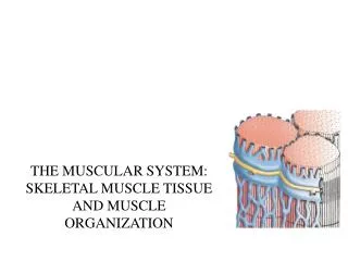
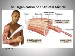
The Organization of a Skeletal Muscle. Figure 7-1. Organization of a Skeletal Muscle Fiber. Figure 7-2a. Organization of a Skeletal Muscle Fiber. Figure 7-2 b-c. Sarcomere Shortening. Figure 7-3. Sarcomere Shortening. Figure 7-3. Structure and Function of the Neuromuscular Junction.

E N D
The Organization of a Skeletal Muscle Figure 7-1
Organization of a Skeletal Muscle Fiber Figure 7-2a
Organization of a Skeletal Muscle Fiber Figure 7-2 b-c
Sarcomere Shortening Figure 7-3
Sarcomere Shortening Figure 7-3
Structure and Function of the Neuromuscular Junction Figure 7-4 a
Structure and Function of the Neuromuscular Junction Figure 7-4 b
Structure and Function of the Neuromuscular Junction Figure 7-4
Structure and Function of the Neuromuscular Junction Figure 7-4
Structure and Function of the Neuromuscular Junction Figure 7-4
Structure and Function of the Neuromuscular Junction Figure 7-4
The Contraction Cycle • Five Steps of the Contraction Cycle • Exposure of active sites • Formation of cross-bridges • Pivoting of myosin heads • Detachment of cross-bridges • Reactivation of myosin
Molecular Events of the Contraction Process The active site is exposed following the binding of calcium ions to troponin. Figure 7-5
Molecular Events of the Contraction Process The myosin cross bridge forms and attaches to the exposed active site on the thin filaments. Figure 7-5
Molecular Events of the Contraction Process The attached myosin head pivots toward the center of the sarcomere and ADP and a phosphate group are released. Figure 7-5
Molecular Events of the Contraction Process The cross-bridges detach when the myosin head binds another ATP molecule Figure 7-5
Molecular Events of the Contraction Process The detached myosin head is reactivated as it splits the ATP and captures the released energy. Figure 7-5
Frequency of Muscle Fiber Stimulation • Three Phases of Twitch • Latent period before contraction: • The action potential moves through sarcolemma • Causing Ca2+ release • Contraction phase: • Calcium ions bind • Tension builds to peak • Relaxation phase: • Ca2+ levels fall • Active sites are covered • Tension falls to resting levels
Twitch and Development of Tension Figure 7-6
Effects of Repeated Stimulations Figure 7-7
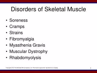
Types of Skeletal Muscle Fibers • Fast fibers (White) • Light in color, large diameter • Few capillaries, few mitochondria • Uses glycolysis (2 ATP) • Little myoglobin • Power/speed • Fatigue rapidly
Types of Skeletal Muscle Fibers • Slow fibers (Red) • Red in color, small diameter • Many capillaries, numerous mitochondria • Uses Krebs cycle/Oxidative phosphorylation (34 ATP) • Much myoglobin • High endurance
Muscle Metabolism Figure 7-9
Muscle Metabolism Figure 7-9
Muscle Metabolism Figure 7-9