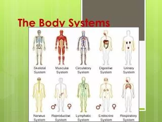
Human Biology: Circulatory System
Human Biology: Circulatory System. Lesson 3: Structure and Function of the Heart (Inquiry into Life pg. 220-226). Today’s Objectives. Describe the inter-relationships of the structures of the heart, including:

Human Biology: Circulatory System
E N D
Presentation Transcript
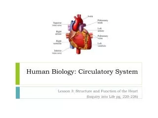
Human Biology: Circulatory System Lesson 3: Structure and Function of the Heart (Inquiry into Life pg. 220-226)
Today’s Objectives • Describe the inter-relationships of the structures of the heart, including: • Identify and give functions (including direction of blood flow) for the major structures of the heart • Recognize heart structures using both internal and extrenal diagram views • Analyse the relationship between heart rate and blood pressure, including: • Describe the location and functions of the sinoatrial (SA) node, atrioventricular (AV) node, and Purkinje fibres • Describe how the autonomic nervous system increases and decreases heart rate and blood pressure • Differentiate between systolic and diastolic pressures • Describe hypertension and hypotension and their causes • Demonstrate the measurement of blood pressure
Parts of the Heart (video) • Left and Right Atria – Collecting Chambers • Right: collects blood from Venae Cavae • Left: Collects blood from Pulmonary Veins • Left and Right Ventricles – Pumps • Right: Sends blood to the lungs via the Pulmonary Trunk • Left: Sends blood to the body via the Aorta
Parts of the Heart • Atrioventricular Valves • Valves between the atria and ventricles • Prevent backflow of blood • Right side valve called “Tricuspid Valve” • Has three cusps, or flaps • Also called right atrioventricular valve • Left side valve called “Bicuspid Valve” • Has two cusps, or flaps • Also know as the mitral valve or left atrioventricular valve • Chordae Tendonae • Strong, fibrous strings that support the A.V. valves • Keep the flaps from inverting with the force of blood flow
Parts of the Heart • Semi-lunar Valves (no chordae tendonae) • Between ventricles and the Aorta (Aortic Valve) • Between the ventricles and the Pulmonary Trunk (Pulmonary Valve) • Pulmonary Trunk • Branches off to form the Pulmonary Artery • Receives blood from the right ventricle • Septum (Ventricular Septum) • The wall of the Heart • Separates the left and right sides of the Heart
Cardiac Cycle and Intrinsic Beat • Contraction of the Heart is a two step process: • Systole – Contraction of the Heart • Diastole – Relaxation of the Heart • Each heart beat (Cardiac Cycle) consists of: TIMEATRIAVENTRICLES 0.15 Sec Systole Diastole 0.30 Sec Diastole Systole 0.40 Sec Diastole Diastole 0.85 Sec • Average time of 70 beats per minute
Cardiac Cycle • The ventricles have a stronger and longer contraction because blood must be pumped throughout the body • The “lub-dup” sound of the heart is due to the closing of the valves: first the atrioventricular, then the semi-lunar • The beat of the heart is said to be intrinsic, meaning it will beat without any nervous system stimulation • It can be removed from the body and still continue beating!) • The beat is controlled by a special type of tissue called Nodal Tissue, which has both muscular and nervous tissue characteristics
Nodal Tissue • There are two locations of Nodal Tissue in the Heart: • 1) SA Node (Sinoatrial Node) • Found in the upper wall of the right atrium • 2) AV Node (Atrioventricular Node) • Found at the bottom of the right atrium near the Septum • The SA Node (also called the pacemaker) initiates the heartbeat and sends out an excitation impulses every 0.85 seconds. • The impulse causes both Atria to contract. The impulses are sent to the AV Node Via the bundle of His. • When the impulse reaches the AV Node, an impulse is sent from the AV Node, down the Purkinje Fibers causing both ventricles to contract.
Electrocardiograms (EKG) • An electrocardiogram registers the voltage change across the surface of the heart as it beats. • The letters PQRST are the standard labels used to identify the parts of the EKG • The P curve records the simultaneous contraction of the atria as they drive the blood out into their ventricles • The QRS is the contraction of the ventricles as they drive the blood out into their respective arteries • The T marks the recovery of the Ventricles (restoration of the normal electrical condition, preparing them for the next contraction)
Autonomic Control of the Heart • The rate of the heart can also be controlled by the nervous system • The heart rate center is located in the Medulla Oblongata of the brain. • The SA Node is connected to the brain by the vagus nerve (cranial nerve #10) • This nerve pathway is called the Autonomic Nervous System (not under conscious control) • Has two systems that affect the Heart Rate: • Parasympathetic System – causes the heart beat to slow down • Sympathetic System – causes the heart beat to increase during times of stress
Autonomic Nervous System • Factors such as a need for oxygen or the blood pressure level determine which of those systems become active • When the brain perceives that the blood is getting delivered to the tissues too slowly, or if blood pressure is low, the brain will signal the SA Node to speed up its contraction.
Blood Pressure • Ventricles pump a volume of blood (approx 70 ml) each time they contract • Must have elastic, expandable walls • The force of blood against the blood vessel walls is known as Blood Pressure • Blood Pressure is not constant • The term Systolic pressure (or Systole) refers to the blood pressure when the ventricles contract • The term Diastolic pressure (Diastole) refers to the blood pressure when the heart is at rest
Pulse • As blood is pumped through the arteries, the arterial walls swell, then relax • This swelling can be felt in any artery that runs close to the surface • Blood pressure is normally measured along the brachial artery of the arm • A reading of 120/80 is quite normal • 120 - Systolic reading as ventricles contract • 80 – Diastolic reading as the heart relaxes
Blood Pressure Changes • A number of things can affect the blood pressure: • Hypertension: High Blood Pressure • Example: 140/90 or 125/90 • Diet and Lifestyle are often to blame for elevated blood pressure • Reasons for Hypertension: • Stress • Plaques – formed by fatty acid deposits from digested foods; line the walls of the arteries, making the radius smaller, thereby raising blood pressure, (Arteriosclerosis, Stroke, Heart Attack) • High Salt Intake – retains water – greater fluid volume leads to greater volume of blood
Blood Pressure Changes • Hypertension continued.. • Smoking • Stimulants • Lack of Exercise • Diet – amount of food and type • Working too hard • Age, Sex, Race
Hypotension • Low Blood Pressure • Blood does not reach all organs • Example: 110/70 • Reasons for Hypotension: • Cuts or amputated limbs • Drugs • Hormones
Effects of Blood Pressure Change • Proper Kidney function can only be maintained if there is a sufficient pressure for filtration • Luckily, the body can adjust blood pressure • Monitored by the Hypothalmus (part of the brain), the body can dilate (widen) arterioles thus lower blood pressure in them, or constrict (narrow) them to raise the blood pressure