END
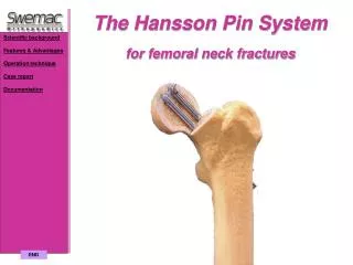
The Hansson Pin System for femoral neck fractures. Scientific background Features & Advantages Operation technique Case report Documentation. END. Scientific background. Scientific background Features & Advantages Operation technique Case report Documentation.

END
E N D
Presentation Transcript
The Hansson Pin System for femoral neck fractures Scientific background Features & Advantages Operation technique Case report Documentation END
Scientific background Scientific background Features & Advantages Operation technique Case report Documentation In 1978 at the University Hospital of Lund in Sweden, a prospective long-term study of pre- and post-operative femoral head vitality and it’s correlation to the clinical and radiographic course was commenced. It was handled by Dr Lars Ingvar Hansson, MD, and his colleagues Dr Göran Bauer, Dr David Weber and Dr Björn Strömqvist.Femoral head vitality was studied with tetracycline before nailing and scintimetry after nailing. The prognostic value of scintimetry was revealed when discrepancies between femoral head vitality before and after nailing was observed. This shows that the four flanged nail (the method of fixation in use at the time) could actually increase the risk of circulatory injury during the operative procedure. Less traumatic methods of fixation were explored and the Hansson pin, which was then in use for slipped capital femoral epiphysis in children, was the result.
Features & Advantages Scientific background Features & Advantages Preserving the blood supply Maximum resistanceto rotation Continuouscompression Minimum disruption to bone Reducing trauma Immediate weight bearing Operation technique Case report Documentation Preserving the blood supply Maximum resistance to rotation Continuous compression Minimum disruption to bone Reducing trauma Immediate weight bearing
Preserving the blood supply Scientific background Features & Advantages Preserving the blood supply Maximum resistanceto rotation Continuouscompression Minimum disruption to bone Reducing trauma Immediate weight bearing Operation technique Case report Documentation The Hansson pin and operative procedure are both deisgned to avoid per-operative trauma. The pins are slid into position into prepared holes which are just oversize. No hammering or turning force is applied to the head when the pins are inserted, thus greatly improving the chance of continued femoral head vitality.
Maximum resistance to rotation Three point support Scientific background Features & Advantages Preserving the blood supply Maximum resistance to rotation Continuouscompression Minimum disruption to bone Reducing trauma Immediate weight bearing Operation technique Case report Documentation Each pin contacts strong cortical bone in three places to provide maximum resistance to rotation. The distal pin prevents varus angulation and adding the proximal pin prevents dorsal angulation of the femoral head.
Continuous compression Parallel placement of the pins Scientific background Features & Advantages Preserving the blood supply Maximum resistanceto rotation Continuous compression Minimum disruption to bone Reducing trauma Immediate weight bearing Operation technique Case report Documentation The two Hansson pins which are used in parallel maximise the natural physiological compressive forces about the hip. With the head and pins fixed together the femoral neck can slide on the pins, giving continuous compression at the fracture site. The drillguide ensures parallel placement of the pins.
Minimum disruption to bone Scientific background Features & Advantages Preserving the blood supply Maximum resistanceto rotation Continuouscompression Minimum disruption to bone Reducing trauma Immediate weight bearing Operation technique Case report Documentation The pin is only 6.5 mm in diameter. I has been proved that too much metal is biologically unfaverable regarding the viability of the femoral head. The Hansson Pins has no additional fixation points in the femoral shaft like the standard compression hip screw thus less risk of a refracture.
Reducing trauma Scientific background Features & Advantages Preserving the blood supply Maximum resistanceto rotation Continuouscompression Minimum disruption to bone Reducing trauma Immediate weight bearing Operation technique Case report Documentation The complete procedure is carried out through a 4-5 cm incision. Simple instrumentation and uncomplicated procedure allow fixation to be achieved within 15 minutes. The procedure lends itself to spinal anaesthesia. The procedure for pin removal is quick and straightforward.
HanssonStudies Immediate weight bearing Scientific background Features & Advantages Preserving the blood supply Maximum resistanceto rotation Continuouscompression Minimum disruption to bone Reducing trauma Immediate weight bearing Operation technique Case report Documentation The Hansson Pin System gives the security and stability of fixation to allow most patient to be mobilised during their first postoperative day and discharged early. Minimising the risks with prolonged bed rest.
Scientific background Features & Advantages Operation technique Reduce fracture Make incision Insert distal guide wire Drill distal channel Select drillguide Insert proximal guidewire Drill proximal channel Remove proximal drill Fix pin to introducerassembly Insert proximal pin Remove distal drill Insert distal pin Close the wound Pin removal Case report Documentation Operation technique
Scientific background Features & Advantages Operation technique Reduce fracture Make incision Insert distal guide wire Drill distal channel Select drillguide Insert proximal guidewire Drill proximal channel Remove proximal drill Fix pin to introducerassembly Insert proximal pin Remove distal drill Insert distal pin Close the wound Pin removal Case report Documentation 1. Reduce fracture Reduction should be obtained by gentle manipulation according to the normal procedure for displaced fractures
Scientific background Features & Advantages Operation technique Reduce fracture Make incision Insert distal guide wire Drill distal channel Select drillguide Insert proximal guidewire Drill proximal channel Remove proximal drill Fix pin to introducerassembly Insert proximal pin Remove distal drill Insert distal pin Close the wound Pin removal Case report Documentation 2. Make incisionA 4-5 cm longitudinal incision is made and the facia lata is divided in the direction of the fibres.
Scientific background Features & Advantages Operation technique Reduce fracture Make incision Insert distal guide wire Drill distal channel Select drillguide Insert proximal guidewire Drill proximal channel Remove proximal drill Fix pin to introducerassembly Insert proximal pin Remove distal drill Insert distal pin Close the wound Pin removal Case report Documentation 3. Insert distal guide wire The guide wire is inserted through the facia. In the AP-view the tip of the guide wire should be leveled with or just below the lower edge of the lesser trochanter. In the lateral view it should be central in relation to the femoral head and neck. It is essential to have the guide wire very close to the inner medial cortex. Once the alignment of the guide wire is satisfactory, it is advanced to the subchondral bone of the femoral head.
Scientific background Features & Advantages Operation technique Reduce fracture Make incision Insert distal guide wire Drill distal channel Select drillguide Insert proximal guidewire Drill proximal channel Remove proximal drill Fix pin to introducerassembly Insert proximal pin Remove distal drill Insert distal pin Close the wound Pin removal Case report Documentation 4. Drill distal channel A cannulated drill and the protective measuring sleeve are inserted over the end of the guide wire. The protective measuring sleeve is pressed against the lateral cortex and the drill is advanced to the subchondral bone of the femoral head. The required length of pin is read off the scale on the drill protruding from the sleeve. The drill is left in position and the protective measuring sleeve is then removed.
Scientific background Features & Advantages Operation technique Reduce fracture Make incision Insert distal guide wire Drill distal channel Select drillguide Insert proximal guidewire Drill proximal channel Remove proximal drill Fix pin to introducerassembly Insert proximal pin Remove distal drill Insert distal pin Close the wound Pin removal Case report Documentation 5. Select drillguide Select a drillguide which gives the widest distance between the two pins without cutting through the posterior cortex (6, 8 or 10 mm). The appropriate selected drillguide is then pushed over the distal drill and rotated, in order that the new channel is situated posteriorly to the distal drill.
Scientific background Features & Advantages Operation technique Reduce fracture Make incision Insert distal guide wire Drill distal channel Select drillguide Insert proximal guide wire Drill proximal channel Remove proximal drill Fix pin to introducerassembly Insert proximal pin Remove distal drill Insert distal pin Close the wound Pin removal Case report Documentation 6. Insert proximal guide wireA small protective sleeve is introduced through the unoccupied hole of the drillguide. The long guide wire is introduced and drilled into the subchondral bone of the femoral head, using image intensification in both AP- and lateral views. The small protective sleeve can then be removed.
Scientific background Features & Advantages Operation technique Reduce fracture Make incision Insert distal guide wire Drill distal channel Select drillguide Insert proximal guidewire Drill proximal channel Remove proximal drill Fix pin to introducerassembly Insert proximal pin Remove distal drill Insert distal pin Close the wound Pin removal Case report Documentation 7. Drill proximal channel A long cannulated drill is introduced through the unoccupied hole of the drillguide and drilled over the guide wire into the subchondral bone of the femoral head. The pin length for the proximal channel is read off against the protruding part of the drill at the end of the drillguide.
Scientific background Features & Advantages Operation technique Reduce fracture Make incision Insert distal guide wire Drill distal channel Select drillguide Insert proximal guidewire Drill proximal channel Remove proximal drill Fix pin to introducerassembly Insert proximal pin Remove distal drill Insert distal pin Close the wound Pin removal Case report Documentation 8. Remove proximal drillThe proximal drill and the drillguide is removed.
Pin Handle Outer introducer Inner introducer Scientific background Features & Advantages Operation technique Reduce fracture Make incision Insert distal guide wire Drill distal channel Select drillguide Insert proximal guidewire Drill proximal channel Remove proximal drill Fix pin to introducer assembly Insert proximal pin Remove distal drill Insert distal pin Close the wound Pin removal Case report Documentation 1. The outer introducer is passed over the inner introducer. 2. The inner introducer is then screwed into the base of the pin. The window for the tongue will be situated on the same side as the mark on the outer introducer and indicates the direction, which the tongue will take when extruded. 3. The tip of the handle is inserted through the channel of the inner introducer and rotated clockwise until it meets resistance, that is, the tip touches the tongue. 9. Fix pin to introducer assembly Very important! The window of the pin should point downwards during assembly.
Guide-line Scientific background Features & Advantages Operation technique Reduce fracture Make incision Insert distal guide wire Drill distal channel Select drillguide Insert proximal guidewire Drill proximal channel Remove proximal drill Fix pin to introducerassembly Insert proximal pin Remove distal drill Insert distal pin Close the wound Pin removal Case report Documentation 10. Insert proximal pin The proximal pin is introduced, ensuring that the guide-line on the outer introducer is pointing anteriorly. When the pin is seen to be in position, the hook is activated by turning the introducer handle clockwise as far as it will go. The introducer assembly is then removed.
Scientific background Features & Advantages Operation technique Reduce fracture Make incision Insert distal guide wire Drill distal channel Select drillguide Insert proximal guidewire Drill proximal channel Remove proximal drill Fix pin to introducerassembly Insert proximal pin Remove distal drill Insert distal pin Close the wound Pin removal Case report Documentation 11. Remove distal drill
Guide-line Scientific background Features & Advantages Operation technique Reduce fracture Make incision Insert distal guide wire Drill distal channel Select drillguide Insert proximal guidewire Drill proximal channel Remove proximal drill Fix pin to introducerassembly Insert proximal pin Remove distal drill Insert distal pin Close the wound Pin removal Case report Documentation 12. Insert distal pin A pin of the length required for the distal channel is mounted on the introducer assembly and inserted under image intensification in the same way, but with the guide-line on the outer introducer facing superiorly. The introducer assembly is then removed.
Scientific background Features & Advantages Operation technique Reduce fracture Make incision Insert distal guide wire Drill distal channel Select drillguide Insert proximal guidewire Drill proximal channel Remove proximal drill Fix pin to introducerassembly Insert proximal pin Remove distal drill Insert distal pin Close the wound Pin removal Case report Documentation 13. Close the wound
Scientific background Features & Advantages Operation technique Reduce fracture Make incision Insert distal guide wire Drill distal channel Select drillguide Insert proximal guidewire Drill proximal channel Remove proximal drill Fix pin to introducerassembly Insert proximal pin Remove distal drill Insert distal pin Close the wound Pin removal Case report Documentation 14. Pin removal A small incision is made for pin removal. The end of the pin can be identified manually or using image intensification. The fibrous tissue which often surrounds the end of the pin is incised. The extractor and the outer introducer are located against the pin, the extractor is then screwed clockwise. This withdraws the hook back into the body of the pin, which can then be removed.
Documentation Scientific background Features & Advantages Operation technique Case report Documentation Theses Publications Theses Publications
Theses Scientific background Features & Advantages Operation technique Case report Documentation Theses Publications 1. Femoral head vitality after intracapsular hip fracture.Björn Strömqvist, 1983. 2. Primary osteosynthesis for femoral neck fracture. Lars T Nilsson, 1989. 3. Femoral neck fracture stability. Evaluation with roentgen sterophotogrammetric analysis, magnetic resonance imaging, scintimetry, radiography and histophatology. Jon Ragnarsson, 1991.
Publications Scientific background Features & Advantages Operation technique Case report Documentation Theses Publications 1. Dynamics of the Technetium-99m methylenediphosphonate imaging of the femoral head after hip fracture. G Bauer, Weber , L Ceder, LI Hansson Clin Orthop 1980 Oct:(152):85-92 2. Vitality of the femoral head after femoral neck fracture evaluated by tetracycline labelling. Strömqvist B, Ceder L, Hansson LI, Thorngren KG Arch Orthop Trauma Surg 99:1-6, 1981 3. 85Sr-scintimetry in femoral neck fracture. Brummer R, Hansson LI, Sjöstrand LO Arch Orthop Trauma Surg 1982:101(1):47-51 4. Nailing of femoral neck fracture can cause vascular injury and segmental collapse. Strömqvist B, Hansson LI Läkartidningen 1982 Feb 24:79(8):659-62 5. Scintimetric evaluation of nailed femoral neck fractures with special reference to type of osteosynthesis. Strömquist B, Hansson LI, Palmer J Acta Orthop Scand 1983 Jun:54(3):340-7 6. Femoral head vitality after femoral neck fracture. Comparison between pre- and peroperative tetracycline labelling. Strömqvist B, Hansson LI Arch Orthop Trauma Surg 1983:101(4):251-7 7. Avascular necrosis associated with nailing of femoral neck fracture. Two cases examined pre and postoperatively by tetracycline and radionuclide tracer techniques. Strömqvist B, Hansson LI Acta Orthop Scand 1983 Oct:57(5):687-94
8. A radiographic five year follow-up of femoral neck farctures. Brummer R, Hansson LI, Mortensson W Acta Orthop Scand 1983 Dec:54(6):895-71 9. Femoral head vitality after intracapsular hip fracture, 490 cases studied by intravital tetracycline labelling and Tc-MDP radionuclide imaging. Strömqvist B Acta Ortop Scand Suppl 200:1-71,1983 10. Emission tomography in femoral neck fracture for evaluation of avascular necrosis. Strömqvist B, Brismar J, Hansson LI Acta Ortop Scand 54:872-7, 1983 11. Technetium-99m-methylendiphosphonate scintimetry after femoral neck fracture. A three-year follow-up study. Strömqvist B, Brismar J, Hansson LI Clin Orthop 1984 Jan-Feb:(182):177-89 12. Femoral head vitality in femoral neck fracture after hook-pin internal fixation. Strömquist B, Hansson LI Clin Ortop 1984 Dec:(191):105-9 13. Two-year follow-up of femoral neck fractures. Comparison of ostesynthesis methods. Strömqvist B, Hansson LI, Nilsson LT Acta Ortop Scand 1984 Oct:55(5):521-5 14. Femoral head vitality at reoperation for femoral neck fracture complications. Strömqvist B, Hansson LI, Palmer J Arch Orthop Trauma Surg 1984:103(4):235-40 15. Hip fracture in rheumatoid arthritis. Strömqvist B Acta Ortop Scand 1984 Dec:55(6):624-8 16. Preoperative 99mTc-MDP scintimetry of femoral neck fractures. Holmberg S, Thorngren KG Acta Orthop Scand 1984 Aug:55(4):430-5 Scientific background Features & Advantages Operation technique Case report Documentation Theses Publications
17. External and biopsy determination of peroperative Tc-99m MDP femoral-head labbeling in fracture of the femoral neck. Strömqvist B, Brismar J, Hansson LI J Nucl Med 1984 Aug:25(8):854-8 18. Pre-operative and postoperative scintimetry after femoral neck fracture. Strömqvist B, Hansson LI, Ljung P J Bone Joint Surg :Br: 1984 Jan:66(1):49-54 19. Evaluation in femoral neck fracture scintimetry: Modes of region interest selection and influence on results. Holmberg S, Mesko L, Strömqvist B J Nucl Med 1985 26:353-6 20. The longest delay between femoral neck fracture and femoral head collapse. Strömqvist B Arch Orthop Trauma Surg 104:125-8 21. Improved operations for femoral neck fracture. A radiographic evaluation. Johansson A, Strömquist B, Bauer G, Hansson LI Acta Orthop Scand 1986 57:505-9 22. Hook-pin fixation in femoral neck fractures. A two-year follow-up study of 300 cases. Strömqvist B, Hansson LI, Nilsson LT Clin Orthop 218:58-62, 1987 23. Effects of strategy changes in the treatment of femoral neck fractures during a 17-year period. Ceder L, Strömqvist B, Hansson LI Clin Orthop 1987 218:53-7 24. Prognostic precision in postoperative, 99Tc-MDP scintimetry after femoral neck fracture. Strömqvist B, Hansson LI, Nilsson LT Acta Ortop Scand 1987 58:494-8 25. Displacement in femoral neck fractures. A numerical analysis of 200 fractures. Eliasson P, Hansson LI, Kärrholm J Acta Orthop Scand 1988 Aug:59(4):361-4 Scientific background Features & Advantages Operation technique Case report Documentation Theses Publications
26. Nailing of femoral neck fracture. Clinical and sociological 5year follow-up of 510 consecutive hips. Nilsson LT, Strömqvist B, Thorngren KG Acta Orthop Scand 1988 Aug:59(4):365-71 27. Hemarthrosis after femoral neck fracturefixation. Egund N, Strömqvist B, Nilsson LT Acta Orthop Scand 1988 Oct:59(5):526-9 28. Treatment of hip fractures in rheumatoid arthritis. Strömqvist B, Kelly I, Lidgren L Clin Orthop 1988 Mar:(228):75-8 29. Fixation of fractures of the femoral neck using screws or hook-pins. Radionnuclide study and short-term results. Strömqvist B, Hansson LI, Ross H Rev Chir Orthop 1988:74(7):609-13 30. Intracapsular pressures in undisplaced fractures of the femoral neck. Strömquist B, Nilsson LT, Egund N J Bone Joint Surg :Br: 1988 Mar:70(2):192-4 31. Internal fixation of femoral neck fractures in Parkinson’s disease. Patients followed for 2 years. Londos E, Nilsson LT, Strömqvist B Acta Orthop Scand 1989 Dec:60(6):682-5 32. Function after hook-pin fixation of femoral neck fractures. Prospective 2-year follow-up pf 191 cases. Nilsson LT, Strömqvist B, Thorngren KG Acta Orthop Scand 1989 Oct:60(5):573-8 33. Secondary arthroplasty for complications of femoral neck fracture. Nilsson LT, Strömqvist B, Thorngren KG J Bone Joint Surg :Br: 1989 Nov:71(5):777-81 34. Stability of femoral neck fractures. A postoperative roentgen stereophotogrammetric analysis. Ragnarsson JI, Hansson LI, Kärrholm J Acta Orthop Scand 1989 Jun:60(3):283-7 Scientific background Features & Advantages Operation technique Case report Documentation Theses Publications
35. CT scans and lipohaemarthrosis in hip fractures. Egund N, Nilsson LT, Wingstrand H J Bone Joint Surg :Br: 1990 May:72(3):379-82 36. Redisplacement of nailed femoral neck fractures. 4-year follow-up of 110 cases. Eliasson P, Kärrholm J, Hansson LI Acta Orthop Scand 1990 Feb:61(1):12-5 37. Secondary total hip replacement after fractures of the hip. Franzen H, Nilsson LT, Strömqvist B J Bone Joint Surg :Br: 1990 Sep:72(5):784-7 38. Measurements of femoral neck fracture stability. Conventional radiography vs. roentgen stereophotogrammetric analysis. Ragnarsson JI, Eliasson P, Kärrholm J Submitted. 39. Stability of femoral neck fractures during weight-bearing. Ragnarsson JI, Kärrholm J Acta Orthop Scand. In press. 40. Factors influencing postoperative movements in displaced femoral neck fractures. Evaluation with conventional and stereo-radiography (RSA). Ragnarsson JI, Kärrholm J Submitted. 41. 626 cases of femoral neck fractures. Nilsson LT, Strömqvist B, Thorngren KG Submitted. 42. Factors predicting healing complications in femoral neck fractures. Nilsson LT, Johansson Å, Strömqvist B Submitted. 43. Function of the hip after femoral neck fractures treated by fixation or secondary total hip replacement. Nilsson LT, Franzen H, Strömqvist B, Wiklund I Int Orthop 1991:(15):315-18 Scientific background Features & Advantages Operation technique Case report Documentation Theses Publications
44. Quality of life is better after osteosynthesis than after hemioarthroplasty in femoral neck fractures. Nilsson LT, Jaalovara P, Franzen H, Virkkunen H, Strömqvist B Submitted. Scientific background Features & Advantages Operation technique Case report Documentation Theses Publications
Case report Scientific background Features & Advantages Operation technique Case report Documentation This is a 34 year old female who fell whilst dancing. She sustained minimal displacement of the head of the femur and no reduction was necessary.These pictures were taken one week after the operation.