Download
1 / 107
1.11k likes | 1.21k Vues
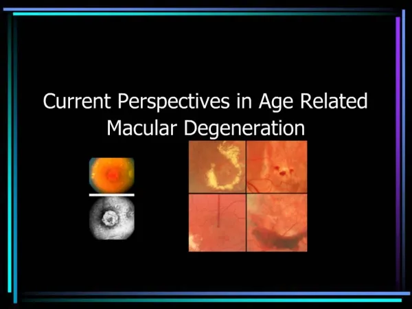
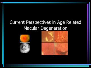
Learn about the progressive eye condition affecting the retina known as Age-related Macular Degeneration (AMD) and its impact on vision. Discover how AMD is diagnosed, stages, symptoms, and potential risk factors.

E N D
How does the eye work? The eye works in much the same way as a camera (figure 1). The cornea, pupil lens and vitreous gel are clear and allow light to focus on a thin sheet of tissue in the back called the retina. The retina itself has photoreceptor cells to transform light energy into nerve impulses as well as nerve fibers to transmit those impulses back to the optic nerve.
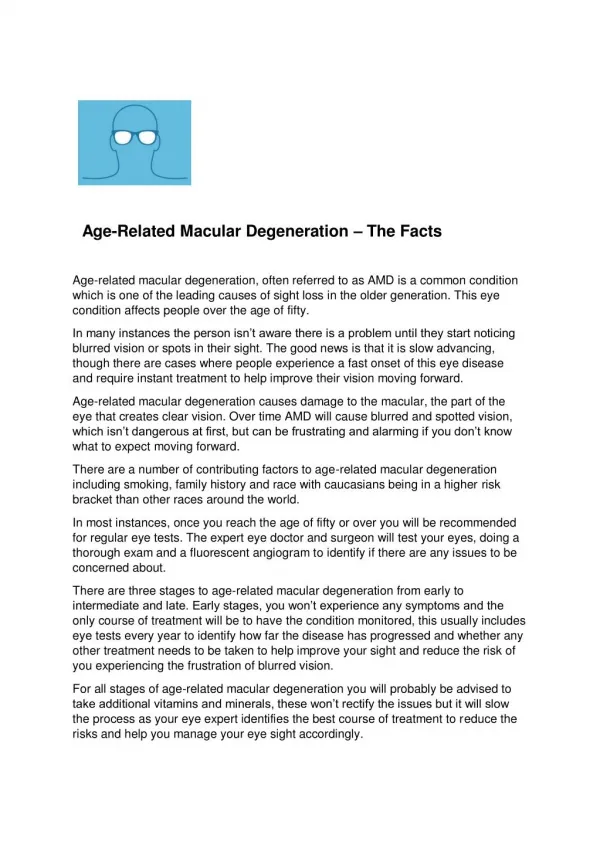
Age-related macular degeneration = eye disease that primarily affects the central portion of the retina known as the macula. The risk for developing macular degeneration increases with age and is in excess of 30% by age 75. The majority of people with macular degeneration have an early form of the condition and experience minimal visual loss. For many of these people, macular degeneration will not progress to a more serious condition. Other risk factors include: a family history of the disease, cigarette smoking, and possibly diet, excessive sunlight exposure, hypertension and cardiovascular disease. Angeles Vision Clinic
Age-related macular degeneration This photograph shows a normal, healthy retina as viewed by an eye doctor during an examination. The macula is the portion of the retina used for detail vision and reading. The center of the macula is called the fovea. Angeles Vision Clinic
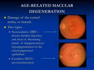
Age-related macular degeneration (AMD) = chronic eye condition that affects age 50 & older. When a person has macular degeneration, the macula begins to deteriorate, causing anything from blurred or slightly distorted central vision to a blind spotin the center of the visual field. Macular degenerationis categorized into stages: dry disease and wet disease. Dry diseaseis the background disease, Wet diseaseis when abnormal blood vessels form as a complication of the dry disease and cause rapid vision loss. Website
Age-related macular degeneration (AMD) How a patient with macular degeneration might see the world. Website
Patchy vision in atrophic macular degeneration How a patient with macular degeneration might see the world.
Distortion of Vision and other symptoms of ARMD Distortion of straight lines which may start to appear crooked over a few weeks usually means the ARMD is progressing. Sometimes this is due to the 'neovascular' ARMD developing, and you are advised to be checked in case laser may help
Age-related Macular Degeneration Disease is associated with an increased Mortality suggesting it is due to a systemic disease
Age-related Macular degeneration => mortality
Age-related Macular Degeneration Disease Pathogenesis
Age-related macular degeneration (AMD) Pathogenesis Website
Early stages of macular degeneration In the early stages of macular degeneration, the transport of nutrients and wastes by the RPE slows down. As waste products accumulate under the retina, they form yellowish deposits calleddrusen. Angeles Vision Clinic
Drusen An ophtalmaologist examining a patient at this stage may note the presence of these drusen, even though most people have no symptoms. When drusen have been noted on examination, monitoring will be needed over time, although most patients will not progress to develop visual loss. Many people over the age of 60 will have some drusen. This retinal photograph shows numerous yellow drusen in & around the macular region of the retina. Angeles Vision Clinic
GeographicAreas of thin retina develop. These areas form like the patterns of countries of the world. The areas get bigger (over years), slowly causing more damage to the sight. Myopic macular degeneration is usually similar.. We now know here that the risk of passive smoking (doubles the risk) & personal smoking (triples the risk) of both geographic atrophy. Localisation of ARMD: 1-2 Very CentralIf the damage is in a very small central area, this may be the type described by Gass, & will not progress.
Localisation of ARMD: 3 • Mixed • Changes may include • thinning of the retina • Drusen • pigmentation, or • thickening of the retina. • There is a variable outcome. ‘Prevention’ may help.
Localisation: 4 Choroidal SclerosisIf there is a large thinned area this may be the ‘choroidal sclerosis’ type of ARMD. This often affects both eyes, but is not treatable, often causing poor central vision. The thin area actually looks white, and the thick choroidal blood vessels can be seen underneath.
Age-related Macular Degeneration Disease Diagnosis
Age-related macular degeneration (AMD):Diagnosis People with macular degeneration may notice rapid onset of symptoms, slight symptoms that progress gradually, or no symptoms at all. Physicians may decide to test for the disease based on family history and any symptoms the patient is experiencing. A thorough eye examination is performed in which the physician looks for abnormalities in the back of the eye, in a portion of the retina called the macula.. Website
Age-related macular degeneration DIAGNOSIS = made based on a thorough eye exam. Eye Exam The doctor looks for presence of abnormalities in the macula, such as deposits called drusen. if the pigmentation is mottled or uneven, instead of its normal even reddish color, macular degeneration is usually the cause. Figure 2. Discrete yellow deposits seen here represent the formation of drusen. Website
Age-related macular degeneration DIAGNOSIS Additional tests are performed to determine the location & extent of the disease: Amsler grid test Fluorescein angiography Indocyanine green angiography (ICGA) Website
Age-related macular degeneration DIAGNOSIS to determine the location & extent of the disease. Amsler grid test As a part of the eye examination, the physician may evaluate the patient's vision using a printed grid. If macular degeneration is present: the lines of the grid may seem faded, broken or distorted. By noting where the distortion occurs (usually near the center of the grid), the doctor can better determine the location & extent of macular damage. Website
How to use The Amsler Grid • 1. Wear your reading eye glasses. • 2. Cover one eye. • 3. Look at the center dot and keep your vision on it at all times. • 4. While looking directly at the central dot and using your peripheral vision: • You should be able to see all four corners. • All the lines should be straight. • All the small squares should be the same size. • Note any distortion. • 5. If you should notice any area on the grid that appears distorted (metamorphopsia), blurred, discolored, or otherwise abnormal (see example to the right), please call your eye care provider right away. • 6. Do this test for each eye.
Small blind spots may appear in your vision as dry macular degeneration progresses. Regular use of the Amsler grid may help you detect any changes in your vision.
Age-related macular degeneration DIAGNOSIS Figure 4. Macular hemorrhage seen in a patient with wet ARMD Note the retinal vessel overlying the hemorrhage. This finding confirms the hemorrhage seen here is subretinal or choroidal in nature. to determine the location and extent of the disease. Fluorescein angiography After diagnosis, the physician may perform this test to determine the extent of the damage from macular degeneration. First, the doctor injects dye into a vein in the patient's arm. As the dye circulates through the bloodstream and eventually to the eye, the blood vessels in the retina stand out as bright yellow when observed with a special blue light. A camera takes flash photographs of the eye every few seconds for several minutes, which help the doctor determine pigmentation changes or abnormal blood vessels.
Age-related macular degeneration DIAGNOSIS to determine the location & extent of the disease. Indocyanine green angiography (ICGA) = Another type of angiography of the vessels in the eye ICG = a dye that lights up when exposed to infrared light. Infrared light is used to take pictures of the back of the eye visualizing retinal blood vessels, & the deeper, harder to see choroidal blood vessels.
Age-related Macular Degeneration Disease Frequency
Macula degeneration In the western world, macular degeneration is the most common cause of severe loss of vision and blindness in persons older than 50. The underlying cause of the condition is a disturbance in the interaction between the retina and choroidof the macula. . In the initial stages, the patient experiences merely amild blurring of vision - Gandorfer A, Haritoglou C, Priglinger S. [Age-related macular degeneration] MMW Fortschr Med. 2005 May 26;147(21):35-8; quiz 39-40. Operative und konservative Retinologie, Intraokulare Chirurgie, Augenklinik der LMU, Munchen. arnd.gandorfer@med.uni-muenchen.de
Dry and wet forms: frequency The dry form represents almost 90% of individuals with ARMD, whereas the wet form is responsible for nearly 90% of severe vision loss.
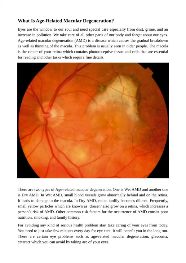
Dry or atrophic ARMD Dry or atrophic form of age-related macular degeneration (ARMD) = progressive condition that begins with the appearance of drusen in the central retinal area. The dry seen above in fundus photography and in a cross-sectional diagram, = the most common form of macular degeneration, accounting for about 90 % of cases. Although this form of AMD does not usually cause severe vision loss, it can progress to the wet form, so patients who have it should see their ophthalmologist regularly. (Cleveland Clinic)
Retinal Pigment Epithelium Atrophy Figure 3. Geographic RPE atrophy results in visualization of the underlying larger choroidal vessels. By Jay M. Haynie, O.D.
Wet , exudative macular degeneration = is usually progressive, = experienced as an acute loss of vision or distortion of the objects viewed. Underlying this wet macular degeneration is of new vessel growth from thechoroid, known as choroidal neovascularization, which as a result of exudationof fluid & bleeding into the macula, destroys central vision. Apart from the vitamin supplementation to slow down progression, laser coagulation, photodynamic treatment or vitreoretinal surgery may be helpful in some cases Gandorfer A, Haritoglou C, Priglinger S. [Age-related macular degeneration] MMW Fortschr Med. 2005 May 26;147(21):35-8; quiz 39-40. Operative und konservative Retinologie, Intraokulare Chirurgie, Augenklinik der LMU, Munchen. arnd.gandorfer@med.uni-muenchen.de
Wet ARMD: Fluorescein Angiography Figure 5. A well-defined focal area of hyperfluorescence seen in the early phase of a fluorescein angiogram in a patient with a classic choroidal neovascularization (CNV). Figure 6. Late phase fluorescein angiogram of a this classic CNV. Note the bright intensity of fluorescence. By Jay M. Haynie, O.D.
Wet ARMD: Fluorescein Angiography Figure 7. Early phase of fluorescein angiogram in an occult CNV. Note the large area of granular fluorescence occupying the entire macula. Figure 8. Late phase fluorescein angiogram of an "occult" CNV. Note the pooling of fluorescein without an identifiable focal source of leakage. By Jay M. Haynie, O.D.
Wet ARMD: Fluorescein Angiography In a minimally classic CNV, fluorescein shows a large neovascular membrane with an area of focal leakage surrounded by a larger area of hypofluorescence or hemorrhage that prevents visualization of the underlying CNV. Figure 9. Late phase of fluorescein angiogram with focal leakage surrounded by dark hypofluorescence. The dark hypofluorescence represents hemorrhage not allowing complete visualization of underlying CNV. hemorrhage By Jay M. Haynie, O.D.
Wet ARMD: Macular hemorrhage Note the retinal vessel overlying the hemorrhage. Figure 4. Macular hemorrhage seen in a patient with wet ARMD This finding confirms the hemorrhage seen here is subretinal or choroidal in nature. By Jay M. Haynie, O.D.
Normal Retina Imagine your retina has five layers. Normally this retinal appearance stays constant even in old age, but changes may develop as you get older.
Exudative types (also called wet) Retinal pigment epithelial detachment (PED): Occult CNV type 1 In a few unlucky people, the dry macular degeneration turns into this type. Occasionally, there will have been no obvious 'dry' changes visible before.In this type the damaged area looks like a dome. Fluid leaks under the retina, hence the term 'wet'.Laser has been tried in this condition but is not helpful, and central vision becomes very poor
Exudative types (also called wet) Classic neovascular ARMD (also called ‘classic CNV’) Some dry types of macular degeneration progress to form this type of wet ARMD. It is very difficult to predict whose dry ARMD will progress, but the risk factors include those mentioned above (soft drusen, high blood pressure, smoking, poor diet). When blood vessels grow under the macula, this is termed choroidal neovascularisation (CNV or CCNV). When the new vessels are seen easily on a fluorescein angiogram, they are called 'classic CNV': They look like a net of blood vessels. New vessels growing under the central retina in a 'classic' pattern: PDT treatment may help
Wet type: Occult’ CNV type 2 (no 'PED') In this type of ARMD, there are new blood vessels, but they are not clearly seen with the angiogram. ‘Occult’ CNV is the term given to a specific ‘blotchy’ appearance of the angiogram. Occult ARMD is probably an early phase of classic.Regular laser is not effective, but even with PDT laser 50% of eyes lose some sight. Occult & classic patterns can occur together. If the % of ‘classic’ is high, PDT helps (early results). New drug treatment probably helps.The symptoms of this type of CNV are the same as 'classic CNV' as above. It often progresses over months or years to cause poor central vision.
Wet (or dry) type: Scarring Many types of macular degeneration progress to cause scarring. 'Dry' types usually progress more slowly, but occasionally can cause very poor central vision, but this is commoner in the 'wet types'.If the patient’s conditions is severe wet, scarring is likely.
What we can we do to prevent, stabilize or slowdown Age-related Macular Degeneration Disease ?