Download
1 / 55
580 likes | 659 Vues
Learn about the increasing prevalence of age-related macular degeneration (AMD) due to ageing population. Explore the causes, types, and effects of this leading cause of severe visual loss. Understand the complex etiology and implications of AMD on central vision.

E N D
An epidemic of “ageing” is impending in the Western world. According to the latest predictions released by the United Nations, the number of people aged over 60 will triple from 606 million worldwide in 2000 to nearly 2 billion by 2050. The increase in population aged over 80 is expected to be more than five fold, from 69 million in 2000 to 379 million by 2050. Ref: BMJ 2003; 326: 485-8
One major implication of this demographic change is the emergence of conditions that are directly related to ageing. Ref: BMJ 2003; 326: 485-8
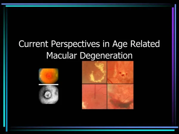
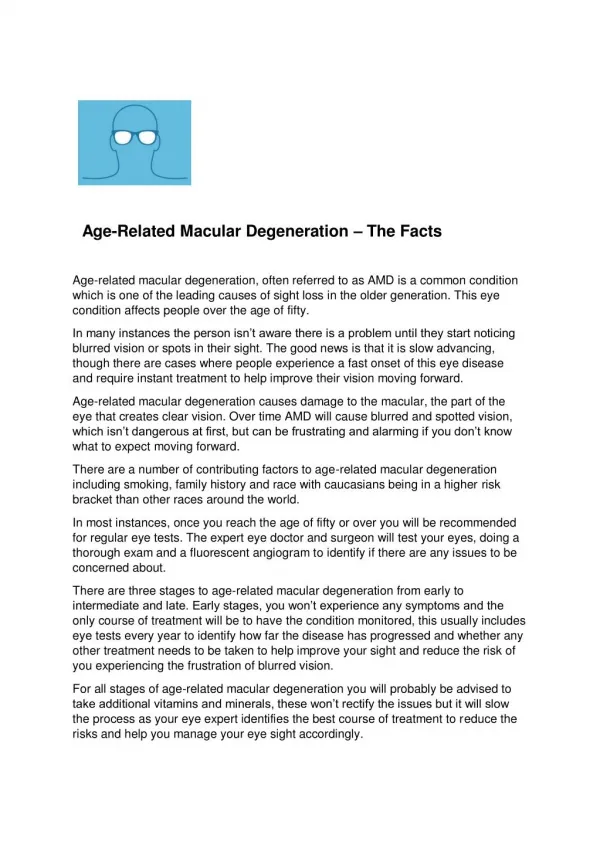
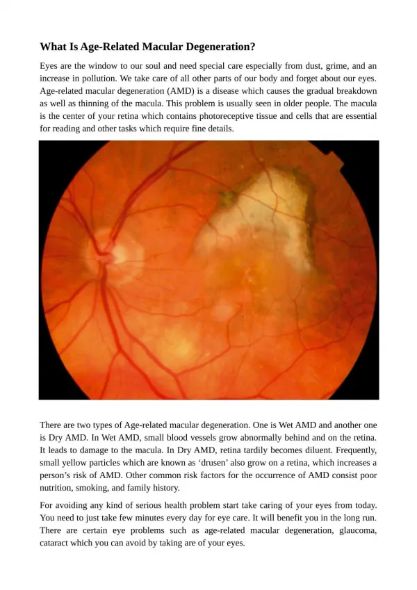
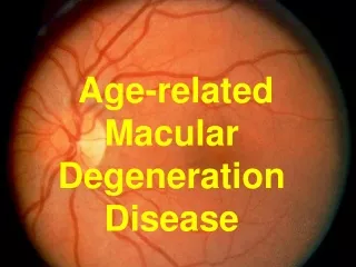
AGERELATED MACULAR DEGENERATION Age related macular degeneration (AMD) is the leading cause of severe visual loss in the western world in people over 50 years of age. Ref: Surveys of ophthalmology 32 (6) 1988: 375-413
AMD: TERMINOLOGY • Degeneration is the change of a tissue to a less functionally active form. • Referred as senile macular degeneration, a name given by Haab as early as 1885, • Age Related macular degeneration has recently been named by Professor A C Bird and coworkers who performed the International ARM Epidemiological study group. • The disorder is either referred to age related maculopathy (ARM) or age related macular degeneration (AMD)
AGE RELATED MACULAR DEGENERATION • The UN estimates the number of people with age related macular degeneration at 20-25 million worldwide • WHO’s estimate is 8 million people with severe visual impairment • AMD was found to be second only to cataract as the cause of severe visual loss Ref: BMJ 2003; 326: 485-8
AMD: PREVALENCE • Prevalence of AMD varies from 1.2% to 29.3% • 3 population based studies; the Beaver Dam Eye Study, Blue Mountain Eye Study and the Rotterdam study report the prevalence rates to be 1.7% in US, 1.4% in Australia and 1.2% in Netherlands respectively • Invest Ophthalmology vis. Sci 2001; 42: 2237-41 • Am J. Epidemiology 1977; 106: 133 • Ophthalmology 1992; 99; 933-43 • Ophthalmology 1995; 102: 1450-60 • Ophthalmology 1995; 102: 205-10
AMD: PREVALENCE IN INDIA In South India, the prevalence is 1.1% whereas, another study from North India reports the prevalence rate to be 4.7% • Invest Ophthalmol Vis Sci (abstract) 2000; 41; 5119 • Ind. J Ophthalmol 1984; 32: 343-6
NORMAL MACULA Ref: http://www/ahaf.org
The macula is the posterior aspect of the retina • Has highest concentration of photoreceptors which facilitate central vision and permit high resolution visual acuity • The macula is an area up to 5.5 mm in diameter with the fovea at its centre NEJM; 342(7): 2000; 483-492
MACULA: CROSS SECTION Ref: http://www.eyesight.org
The retinal pigment epithelium (RPE) is a single layer of hexagonally shaped cells. They reach out to the photoreceptor layer of the retina • Functions of RPE includes maintainance of the photoreceptors, absorption of stray light, formation of the outer blood retinal barrier, phagocytosis and regeneration of visual pigment • Bruch’s membrane separates the RPE from vascular choroid, • Function of Bruch’s membrane is to provide support to the retina • Choroid capillaries are a layer of fine blood vessels that nourishes the retina and provides O2 Ref: http://www.ahaf.org http://www.eyesight.org
Vision in the retina depends on photoreceptor cells (rods and cones) Photoreceptor sit on a layer of RPE Contain pigment called Rhodopsin Opsin->glycoprotein Rhodopsin Cis-retinal -> derivative of vit A Cis-retinal in presence of light Trans-retinal Electric impulse destined for the brain is generated
Also, trans-retinal -> recycled to cis-retinal in RPE. This entire process requires oxygen and nutrition supplied by the fine blood vessels of the choriocapillaries
AMD : Etiology • Etiology is complex and poorly understood • Flawed transport between choroid vessels and photoreceptors may be involved • Angiogenesis is likely to be an early feature of neovascular ARMD
AGE RELATED MACULAR DEGENERATION Insufficient oxygen and nutrients damages photoreceptor molecules With ageing, the ability of RPE cells to digest these molecules decreases Excessive accumulation of residual bodies (drusen) RPE membrane and cells degenerate and atrophy sets in and central vision is lost BMJ 326, 2003; 485-488
AGE RELATED MACULAR DEGENERATION Alternatively the photoreceptors and pigment epithelium send a distress signal to choriocapillaries to make new vessels New vessels grow behind the macula Breakdown in the Bruch’s membrane Blood vessels are fragile Leak blood and fluid Scarring of macula Potential for rapid severe damage BMJ 326; 2003: 485-488
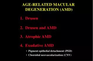
AGE RELATED MACULAR DEGENERATION TYPES • Dry macular degeneration • Wet macular degeneration Ref: NEJM, Vol. 342 (7): 483-492
DRY MACULAR DEGENERATION • Accounts for about 90% of all cases • Also called atrophic, non exudative or drusenoid macular degeneration
DRY MACULAR DEGENERATION Drusen • Drusen is an aggregation of hyaline material located between Bruch’s membrane and RPE • Drusen are composed of waste products from photoreceptors • Drusen > 63 microns in diameter are statistically associated with visual pathology and are termed early ARMD • Hypo/hyper pigmentation of RPE may be present NEJM, Vol 342 (7): 483-492
WET MACULAR DEGENERATION • Accounts for about 10% • Also called choroidal neovascularization, subretinal neovascularization or disciform degeneration • Abnormal blood vessels grow beneath the macula • These vessels leak blood and fluid into the macula damaging photo receptors • Progresses rapidly and can cause severe damage to central vision http://www.blindness.org
AMD: COURSE AND VISUAL PROGNOSIS • Patients with only drusen (in one or both eyes) typically do not have much loss of vision, but they make require additional magnification of the text and more intense lighting to read small point • Presence of large drusen (> 63 microns in diameter) is associated with a risk of the late form of the disease • Patients with large drusen are at relatively high risk for choroidal neovascularization (CNV)
AMD: COURSE AND VISUAL PROGNOSIS Geographic atrophy is the severest form of the dry macular degeneration representing a zone of RPE atrophy 175 microns or greater in diameter with exposure of the underlying choroidal vessels • Leakage of blood or serum as a result of choroidal neovascularization may occur precipitously and is often associated with the abrupt loss of vision • Patients with CNV have a rapid decline in vision (20/200) within weeks • More frequently, visual acuity deteriorates more slowly and stabilises within 3 years • Once CNV has developed in one eye, the other eye is at relatively high risk for the same change NEJM; Vol. 342(7); 483-492
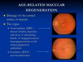
AMD: SYMPTOMS Initial symptoms • Blurry vision • Distorted vision • Straight lines appear wavy • Objects may appear as the wrong shape or size • A dark empty area in the centre of vision http://www.kellogg.umich.edu/
AMD: SYMPTOMS Patient’s ability to perform normal daily tasks such as reading, sewing, telling the time, driving are greatly impaired. http://www.medscape.com
AMD: ESTABLISHED AND POSSIBLE RISK FACTORS NEJM; Vol. 342 (7): 2000: 483-492
AMD: DIAGNOSIS • Visual acuity is tested using the standard eye chart. It measures vision at various distances and can detect vision loss • Amsler grid test: Assesses distorted or reduced vision and small irregularities in the central field of vision • Retinal examination: Done through slit lamp microscope examination: to detect drusen, as well as neovascularization • Fluoroscein angiography: Determines the presence and location of neovascularization Ref: http://www.visionchannel.net
DRY AMD: MANAGEMENT • Low vision aids • Antioxidants
AREDS STUDY Aim To evaluate the effect of anti-oxidant vitamins and zinc on the progression of dry AMD. The study was initiated by National Institutes of Health. No. of centres 11 No. of people 4767 participants aged 55-80 years
AREDS STUDY (contd.) Patients divided into 4 categories: Category 1: little or no AMD -> randomized to antioxidants or placebo to determine any effect on lens changes Category 2: early AMD Category 3: intermediate AMD Category 4: advanced AMD in one eye Category 2, 3 and 4 randomized to receive: • Placebo • Antioxidants alone • Zinc alone • Antioxidants plus zinc (Vit. C: 500 mg, Vit. E: 400 IU, Betacarotene: 15 mg, Zn oxide: 80 mg, Copper: 2mg) Category 2, 3, 4 were followed for visual loss for the development of advanced AMD Patients followed up: 6.3 years
AREDS STUDY (contd.) Results For category 2, only 13% of patients progressed to advanced AMD. For categories 3 and 4 (who are at greater risk for developing advanced AMD), it was found that the combination of zinc and antioxidants were most effective in reducing the progression to advanced AMD. Conclusion It was recommended that patients with intermediate or advanced AMD should consider taking antioxidant vitamins and zinc
WET AMD: MANAGEMENT • Laser photocoagulation • Photodynamic therapy
LASER PHOTOCOAGULATION • Intravenous fluoroscein angiography is performed • Well-circumscribed new blood vessels identified on the fluoroscein angiogram • Treated with laser photo coagulation after topical or local anaesthesia
The Principle of Photodynamic therapy • In contrast with the conventional hot laser • PDT helps to selectively close off subretinal new vessels • two stage treatment • Injecting the photosensitiser drug • Applying cold laser to activate the drug • Releases the singlet oxygen molecule that damages the endothelium • Thrombosis of the capillaries
PHOTODYNAMIC THERAPY • PDT for AMD is a two stage process comprising a 10 minute intravenous infusion of 6 mg/kg verteporfin followed by activation 5 minutes later by 689 nm diode laser for 83 seconds at 503/cm2 • The photosensitive verteporfin is selectively taken up by rapidly proliferating endothelial cells within the target CNV reaching its peak concentration at 15 minutes • Cytotoxic reactive oxygen intermediates damage cellular proteins and cause microvascular thrombosis
PHOTODYNAMIC THERAPY (contd.) • The recent publication of the Treatment of Age-related Macular Degeneration (TAP) report and Verteporfin in Photodynamic Therapy (VIP) trials • For predominantly classic lesions the frequency of stable/improved vision was: 12 months-67% treated, 39% placebo