Glandular Tissue
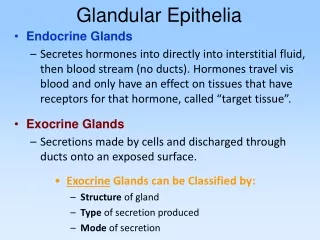
Glandular Tissue. Glands. structures that secrete either onto the surface of a structure or into a lumen of an organ via ducts = exocrine e.g. salivary, lacrimal, sudoriferous or directly into the bloodstream (no ducts) = endocrine e.g. thyroid, pituitary, adrenals. Exocrine Glands.

Glandular Tissue
E N D
Presentation Transcript
Glands • structures that secrete • either onto the surface of a structure or into a lumen of an organ via ducts = exocrine • e.g. salivary, lacrimal, sudoriferous • or directly into the bloodstream (no ducts) = endocrine • e.g. thyroid, pituitary, adrenals
Exocrine Glands • exocrine secretions: • 1. perspiration • 2. digestive enzymes • 3. milk • exocrine gland types: • 1. serous - watery fluid that contains enzymes • e.g. saliva • 2. mucous - glycoproteins called mucins that absorb water to form a • slippery mucus • 3. mixed - more than one type of gland cell • -produces different types of secretions - mucus and serous • e.g. submandibular gland
Glands of the Skin • Specialized exocrine glands found in dermis • Sebaceous (oil) glands • Sudiferous (sweat) glands • Ceruminous (wax) glands • Mammary (milk) glands
Sebaceous (oil) glands • Secretory portion in the dermis • Most open onto hair shafts • Sebum • combination of cholesterol, proteins, fats & salts • keeps hair and skin from soft & pliable • inhibits growth of bacteria & fungi(ringworm) • Acne • bacterial inflammation of glands • secretions stimulated by hormones at puberty
Sudoriferous (sweat) glands • Eccrine (sweat) glands • most areas of skin • secretory portion in dermis with duct to surface • regulate body temperature with perspiration • Apocrine (sweat) glands • armpit and pubic region • secretory portion in dermis with duct that opens onto hair follicle • secretions more viscous
Lacrimal Glands • secrete tears or lacrimal fluid • forms a “tear film” over the surface of the eye – moistens & protects the anterior surface of the eyeball • located in the supero-lateral region of the orbit – in the lacrimal fossa of the frontal bone
lacrimal fluid = tears • each gland drains into 6 • to 12 excretory lacrimal ducts • empties onto the conjuctiva • drains into the lacrimal • puncta -> lacrimal canal -> • lacrimal sac -> naslacrimal • duct -> nasal cavity • -innervated by preganglionic • parasymp. fibers from the greater petrosal nerve • (branch of VII) – synapse with postganglionic fibers of the trigeminal (V) at the pterygopalatine ganglion • -blood supply from the lacrimal and opthalmic arteries Lacrimal Apparatus • About 1 ml of tears produced per day. Spread over eye by blinking. Contains bactericidal enzyme called lysozyme.
Salivary Glands • produce saliva • controlled by the ANS • major and minor glands – defined by size • parotid • submandibular • sublingual • minor glands – • Facial nerve • buccal, labial and lingual mucosal glands • soft palate, hard palate, floor or mouth • Ebner’s glands – associated with the circumvallate papillae/taste buds (secretion of a serous fluid) • secretion of mainly a mucous saliva containing carbohydrates
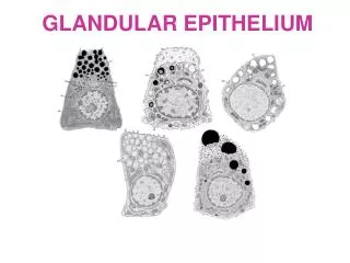
Salivary Gland Cellular Structure • Cells in acini (clusters) • Serous cells secrete a watery fluid • Mucous cells (pale staining) secrete a slimy, mucus secretion
Parotid Gland • occupies the parotid fascial space • innervated by the parasymp. fibers of the otic ganglion of IX + sensory branches of the auriculotemporal branch of V • lymphatic drainage by deep parotid lymph nodes • blood supply by the external carotid artery branches • Parotid below your ear and over the masseter • largest of the major glands • only 25% of total salivary volume – serous fluid • two lobes: superficial and deep • drained by the parotid or Stensen’s duct (superficial to the masseter and pierces the buccinator) • duct opens into the oral cavity opposite the second maxillary molar • parotid papilla (figure 7-5)
Mumps • Myxovirus that attacks the parotid gland • Symptoms • inflammation and enlargement of the parotid • fever, malaise & sour throat (especially swallowing sour foods) • swelling on one or both sides • Sterility rarely possible in males with testicular involvement (only one side involved) • Vaccine available since 1967
Submandibular Gland • provides 60 – 65% of salivary volume • mixed secretion of both serous and mucous • innervated by efferent (parasymp) fibers of chorda tympani and submandibular ganglion of VII • lymphatics drained by submandibular lymph nodes • blood supply by branches of the facial and lingual arteries • submandibular or Wharton’s duct • duct has a very tortuous path over the anterior floor of the mouth • often associated with the formation of salivary stones • removal of these stones can damage the lingual branch of V which is in proximity to the duct • opens into the oral cavity through the sublingual caruncle • near the midline of the mouth floor on each side of the lingual frenulum – figure 7-5 • occupies the submandibular fossa in the submandibular fascial space • most is superficial to the mylohoid muscle
Sublingual Gland • provides on 10% of salivary volume • mix of both serous and mucous secretions • carbohydrate-rich mucous secretion dominates • several short ducts that combine to form the sublingual or Bartholin’s duct • major opening into the oral cavity is the sublingual caruncle • other small ducts open via the sublingual fold – a fold of tissue on the lateral sides of the floor mouth • located in the sublingual fossa in the sublingual fascial space • superior to mylohyoid and anterior to the submandibular gland • innervated by efferent (parasymp) fibers of the chorda tympani and submandibular ganglion of VII • lymphatic drainage into submandibular lymph nodes • blood supply by sublingual and submental branches
Composition and Functions of Saliva • Wet food for easier swallowing • Dissolves food for tasting • Bicarbonate ions buffer acidic foods • bulemia---vomiting hurts the enamel on your teeth • Chemical digestion of starch begins with enzyme (salivary amylase) • Enzyme (lysozyme) ---helps destroy bacteria • Protects mouth from infection with its rinsing action---1 to 1 and 1/2qts/day
Salivation • Increase salivation • sight, smell, sounds, memory of food, tongue stimulation---rock in mouth • cerebral cortex signals the salivatory nuclei in brainstem---(CN 7 & 9) • parasympathetic nn. (CN 7 & 9) • Stop salivation • dry mouth when you are afraid • sympathetic nerves
Thyroid Gland • comprised of microscopic sacs called follicles = follicular cells making up the walls, surrounds a lumen • synthesize T3 & T4 (thyroxin) • In between follicular cells cells are parafollicular cells • produce calcitonin • On each side of trachea is lobe of thyroid • connected by an isthmus • Weighs 1 oz & has rich blood supply
Actions of Thyroid Hormones • T3 & T4 = increases metabolic rate stimulates synthesis of protein stimulates breakdown of fats stimulates cholesterol excretion stimulates synthesis of proteins increases use of glucose & oxygen (ATP production) increases body temperature (calorigenic effect)
Control of T3 & T4 Secretion • Low blood levels of hormones stimulate hypothalamus -> TRH • It stimulates pituitary to release TSH • TSH stimulates gland to raise blood levels • T3 and T4 regulate themselves through a negative feedback loop
Parathyroid Glands • 4 pea-sized glands found on back of thyroid gland • Principal cells produce parathyroid hormone (PTH) • Oxyphil cell function is unknown
Parathyroid Hormone • Raises blood calcium levels • increases activity of osteoclasts • increases reabsorption of Ca+2 by kidney • promote formation of calcitriol (vitamin D3) by kidney which increases absorption of Ca+2 and Mg+2 by intestinal tract • Opposite function of calcitonin (thyroid) • High or low blood levels of Ca+2 stimulate the release of different hormones --- PTH or CT • high level of calcium in blood - release of calcitonin by parafollicular cells, promotes uptake of calcium into bone matrix, lowers blood calcium • low level of calcium in blood - release of PTH by parathyroid glands, promotes release of calcium from bone, raises blood calcium
Thymus Gland • Important role in maturation of T cells • Hormones produced by gland promote the proliferation & maturation of T cells • thymosin • thymic humoral factor • thymic factor • thymopoietin