Download
1 / 1
10 likes | 155 Vues
M. D. A NDERSON Cancer Center. Magnetic Resonance Imaging: 4.7 T, 40 cm Bruker Biospec James A Bankson, Ph.D. and John D Hazle, Ph.D. Small Animal Cancer Imaging Research Facility.

E N D
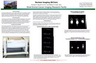
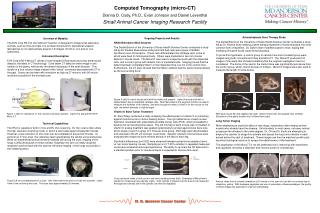
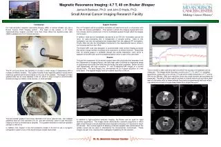
M. D. ANDERSON Cancer Center Magnetic Resonance Imaging: 4.7 T, 40 cm Bruker Biospec James A Bankson, Ph.D. and John D Hazle, Ph.D. Small Animal Cancer Imaging Research Facility Tumor volume is often used as a metric by which the success of a treatment can be measured. The images below illustrate a routine set of T1, T2, and T1+C weighted acquisitions, along with a four minute 3-D scan which yields a resolution of 117 mm by 234 mm by 468 mm. With such resolution, even very small volumes can accurately be measured. The ability to follow specific animals in a serial manner can provide a great deal of information with far fewer animals than previous experimental methods where one animal would yield a single measurement at a single time point. Introduction For high-resolution magnetic resonance imaging (MRI) of animal models, we use a Bruker Avance 47/40 imaging system. The heart of the system, a 4.7 Tesla superconducting magnet, provides more than three times the signal-to-noise ratio (SNR) available from 1.5 T clinical scanners. The 40 cm bore allows the flexibility to conduct a wide range of experiments with a number of different models. The system includes three separate gradient inserts which maximize gradient performance based on the size of the sample. The largest of these gradient sets has an inner diameter of 26 cm, which is large enough to accommodate animals as large as rabbits, small canines, or small primates. The next smaller gradient insert (inner diameter of 12 cm) is ideal for rats. Our smallest gradients have an inner diameter of 6 cm, and are commonly used for high resolution imaging of mice or excised tissue samples. This gradient achieves a maximum gradient strength of 950 mT/m. In addition, this magnet is the first production model of its kind to use a cryogenic refrigeration system in lieu of the liquid nitrogen cooled heat shield. Support Facilities The 4.7T magnet and its acquisition computer and electronics work in conjunction with an SGI O2 console workstation. The operator controls the imaging experiments from this console, which is positioned in front of a shielded window through which the magnet can be seen. In addition, data can be immediately transferred to an SGI O2+ workstation across the room, for data processing that is independent of console activity. Data on this workstation can also be accessed and manipulated from the adjacent PC workstation. Finally, data can also be immediately transferred to the departmental server, where it can be accessed from any office. The Bruker MRI suite was designed to accommodate small animal imaging protocols that call for a large number of animals to be scanned in an efficient manner. Toward that end, full animal support is available through the animal preparation room which is adjacent to the MR suite, and the anesthesia station which is part of the suite itself. Results Through the cooperation of the animal support team with physicists and engineers from the Department of Imaging Physics, we have been able to achieve an impressive range of acquisitions during the brief time that the facility has been underway. Below, left and right respectively, are high resolution T1- and T2-weighted MR images of a normal mouse brain. The axial images, top, demonstrate 49 mm in-plane resolution of 1-mm thick slices. The sagittal images have an in-plane resolution of 59 mm. In addition to high-resolution anatomic imaging, the Bruker can be used for rapid dynamic acquisitions. The images below demonstrate a dynamic acquisition in which eight slices through a mouse abdomen were acquired once every 6.4 seconds for about 35 minutes. Two contrast agents of differing molecular weights were administered; the uptake curves can be used to characterize the microvascular environment. These images are part of an ongoing U54 investigation headed by Dr. Ed Jackson. A H