Excitable Tissues- Synapse
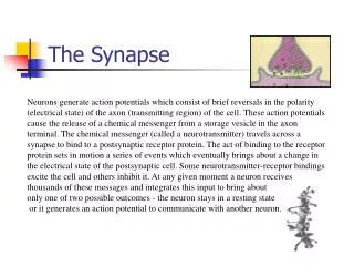
Excitable Tissues- Synapse. Prof. K. Sivapalan. Synapses. Two or more neurons communicate through synapse. Communications are formed with the cell body, dendrides or with the axon sometimes. Structure of a synapse. Pre-synaptic and post synaptic membrane Synaptic cleft- 20-40 nm.


Excitable Tissues- Synapse
E N D
Presentation Transcript
Excitable Tissues- Synapse Prof. K. Sivapalan
Synapses • Two or more neurons communicate through synapse. • Communications are formed with the cell body, dendrides or with the axon sometimes. Synapse
Structure of a synapse • Pre-synaptic and post synaptic membrane • Synaptic cleft- 20-40 nm. • Synaptic vesicles- transmitter • Transmitters released by exocytosis and recovered by endocytosis or destroyed by enzymes Synapse
Sequence of Events at the Axon Terminal • Action potential arrives in the axon • Voltage gated calcium channels at the pre-synaptic membrane open and calcium ions enter • Calcium activates release of synaptic vesicles • Synaptic vesicles cross the cleft and bind with the receptors in the post synaptic membrane. Synapse
Events at the Post Synaptic Membrane • The transmitters released in the cleft bind with receptors in the post synaptic membrane • The transmitter may increase the conductivity (permeability) of cl- or Na+ [sometimes Ca++] • Ligand gated channels • This leads to entry of the respective ion into the ICF resulting in change in membrane potential. • Entry of cation alters the potential towards threshold level [EPSP] and anion causes hyper polarization [IPSP] • EPSP and IPSP from multiple synapses [spacial] or repeated [temporal] release from the same synapse are summated. Synapse
Spatial Summation Synapse
Temporal Summation Synapse
Properties of Dendrites When EPSP or IPSP is generated at a synapse, it affects the potential across all dendrites and the cell body by flow of electrons. Since the axon hillock is the place with lowest threshold, it fires action potential. As the action potential goes away in axon, it also spreads back words along the cell membrane and dendrites wiping out all remnent PSPs refreshing the cell. Synapse
Synaptic Inhibition • Presynaptic Inhibition • Post synaptic inhibition. Synapse
About Synapses • Synaptic delay- 5ms • Transmitters are removed by enzymatic degradation or re-uptake for reuse • Slow PSP with latency of about 100-500 mslasting for several seconds is found in autonomic ganglia, cardiac and smooth muscles and some cortical neurons • As the dendrites branch extensively and have many synapses, the total sum of the PSP will determine genesis of Action Potential • Changes in dendritic spines have been implicated in motivation, learning, and long-term memory. Synapse