Download
1 / 35
410 likes | 938 Vues
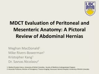
MDCT Evaluation of Peritoneal and Mesenteric Anatomy: A Pictoral Review of Abdominal Hernias. Meghan MacDonald 1 Mike Rivers-Bowerman 1 Kristopher Kang 1 Dr. Savvas Nicolaou 2. 1: Medical Student Intern, University of British Columbia , Faculty of Medicine Undergraduate Program

E N D
MDCT Evaluation of Peritoneal and Mesenteric Anatomy: A Pictoral Review of Abdominal Hernias Meghan MacDonald1 Mike Rivers-Bowerman1 Kristopher Kang1 Dr. Savvas Nicolaou2 1: Medical Student Intern, University of British Columbia , Faculty of Medicine Undergraduate Program 2: Associate Professor, Director of Emergency / Trauma Imaging, Vancouver General Hospital, University of British Columbia
Outline • Definition • Predisposing factors • Clinical presentation • Common types of hernias • Clinical presentation • Utility of MDCT in diagnosis • MDCT features of hernias • Case examples • Conclusion • References
Definition Abdominal hernias include: • Internal hernias (10%): • Involves the protrusion of the bowel through the omentum or mesentery and into a compartment in the abdominal cavity1-4 • Abdominal wall hernias (90%): • Prolapse of an intestinal loop, omentum, or mesentery through a defect in the wall of the abdomen and/or pelvis2-4 • Diaphragmatic hernias (not discussed) Takeyama N, Gokan T, Ohgiya Y, Satoh S, Hashizume T, Hataya K, Kushiro H, Nakanishi M, Kusano M, Munechika H. CT of internal hernias. RadioGraphics 2005;25:997-1015. Strange CD, Birkemeier KL, Sincleair ST, Shepherd JR. Atypical abdominal hernias in the emergency department: acute and non-acute. Am Soc Emergency Radiol 2009;16:121-128. Martin LC, Merkle EM, Thompson WM. Review of internal hernias: Radiographic and clinical findings. AJR 2006;186:703-717. Lee HK, Park SJ, Yi BH. Multidetector CT reveals diverse variety of abdominal hernias. Diagnostic Imaging Asia Pacific [Internet]. 2010 Apr 8 [cited 2010 Aug 1]:1-6. Available from: http://www.diagnosticimaging.com/ct/content/article/113619/1551482
Predisposing Factors Hernial orifices may be normal openings (i.e. hiatal hernia) or may result from: • Congenital defects • Surgery • Trauma • Inflammation • Circulatory problems2,3 2. Strange CD, Birkemeier KL, Sincleair ST, Shepherd JR. Atypical abdominal hernias in the emergency department: acute and non-acute. Am Soc Emergency Radiol 2009;16:121-128. 3. Martin LC, Merkle EM, Thompson WM. Review of internal hernias: Radiographic and clinical findings. AJR 2006;186:703-717.
Clinical Presentation • Most common presentation of abdominal hernias is small bowel obstruction1-4 • Common symptoms include: • Abdominal pain—vague epigastric pain to intermittent colicky periumbilical pain3,5 • Distension • Nausea • Vomiting • Constipation5 • Hernias are the second most common cause of small bowel obstruction2 Takeyama N, Gokan T, Ohgiya Y, Satoh S, Hashizume T, Hataya K, Kushiro H, Nakanishi M, Kusano M, Munechika H. CT of internal hernias. RadioGraphics 2005;25:997-1015. Strange CD, Birkemeier KL, Sincleair ST, Shepherd JR. Atypical abdominal hernias in the emergency department: acute and non-acute. Am Soc Emergency Radiol 2009;16:121-128. Martin LC, Merkle EM, Thompson WM. Review of internal hernias: Radiographic and clinical findings. AJR 2006;186:703-717. Lee HK, Park SJ, Yi BH. Multidetector CT reveals diverse variety of abdominal hernias. Diagnostic Imaging Asia Pacific [Internet]. 2010 Apr 8 [cited 2010 Aug 1]:1-6. Available from: http://www.diagnosticimaging.com/ct/content/article/113619/1551482 Maxhimer JB, Quiros RM. Small bowel obstruction. Common Surgical Diseases 2008;V:141-143.
Utility of MDCT in Diagnosis • First-line imaging technique • Advantages: • Speed • Availability • Multiplanarreformating capabilitiesmake it possible to identify hernias and their complications, including small bowel obstruction and strangulation1,2 • Sensitivity approaches 100%2 Takeyama N, Gokan T, Ohgiya Y, Satoh S, Hashizume T, Hataya K, Kushiro H, Nakanishi M, Kusano M, Munechika H. CT of internal hernias. RadioGraphics 2005;25:997-1015. Strange CD, Birkemeier KL, Sincleair ST, Shepherd JR. Atypical abdominal hernias in the emergency department: acute and non-acute. Am Soc Emergency Radiol 2009;16:121-128.
MDCT Findings of Hernias • Evidence of obstruction with segmental dilatation and stasis (non-specific) 1-8 • Abnormal location of bowel loops3 • “Small bowel feces sign” 2,8 • Gas mixed with particulate in distended small bowel Takeyama N, Gokan T, Ohgiya Y, Satoh S, Hashizume T, Hataya K, Kushiro H, Nakanishi M, Kusano M, Munechika H. CT of internal hernias. RadioGraphics 2005;25:997-1015. Strange CD, Birkemeier KL, Sincleair ST, Shepherd JR. Atypical abdominal hernias in the emergency department: acute and non-acute. Am Soc Emergency Radiol 2009;16:121-128. Martin LC, Merkle EM, Thompson WM. Review of internal hernias: Radiographic and clinical findings. AJR 2006;186:703-717. Lee HK, Park SJ, Yi BH. Multidetector CT reveals diverse variety of abdominal hernias. Diagnostic Imaging Asia Pacific [Internet]. 2010 Apr 8 [cited 2010 Aug 1]:1-6. Available from: http://www.diagnosticimaging.com/ct/content/article/113619/1551482 Maxhimer JB, Quiros RM. Small bowel obstruction. Common Surgical Diseases 2008;V:141-143. Aguirre DA, Casola G, Sirlin C. Abdominal wall hernias: MDCT findings. AJR 2004;183:681-690. Lockhart ME, Tessler FN, Canon CL, Smith JK, Larrison MC, Fineberg NS, Roy BP, Clements RH. Internal hernia after gastric bypass: Sensitivity and specificity of seven CT signs with surgical correlation and controls. AJR 2007;188:745-750. Geraghty P, Desser TS. MDCT of the small bowel. Applied Radiology 2006;35(9): 11-23.
MDCT Findings of Hernias • “Zone of transition” 2,8 • Bowel caliber changes from dilated to a normal or collapsed luminal diameter • 10% of hernias closed loop obstructions with transition zones at the entrance and exit of the hernia sac and dilated bowel between these two sites • Mesenteric vessel engorgement, crowding, twisting, stretching3 • Other findings suggesting strangulation2 • Engorged vessels in an area of incarceration • Increased density in surrounding fat 2. Strange CD, Birkemeier KL, Sincleair ST, Shepherd JR. Atypical abdominal hernias in the emergency department: acute and non-acute. Am Soc Emergency Radiol 2009;16:121-128. 8. Geraghty P, Desser TS. MDCT of the small bowel. Applied Radiology 2006;35(9): 11-23.
CASE STUDIES Internal Hernias
Common Locations of Internal Hernias • A: Foramen of Winslow (8%) • B and C: Left and right paraduodenal (53%) • D: Transmesenteric (8%) • E: Pericecal (13%) • F: Transomental (1-4%) • G: Intersigmoid (6%) Numbers in brackets represent relative frequencies Taken with permission from Reference 1. Takeyama N, Gokan T, Ohgiya Y, Satoh S, Hashizume T, Hataya K, Kushiro H, Nakanishi M, Kusano M, Munechika H. CT of internal hernias. RadioGraphics 2005;25:997-1015.
Common Locations of Internal Hernias • H: Supravesical • I: Broad Ligament • 1: Vesicouterine • 2: Douglas pouch • 3: Perirectal fossa (both total 6%) Numbers in brackets represent relative frequencies Taken with permission from Reference 1. Takeyama N, Gokan T, Ohgiya Y, Satoh S, Hashizume T, Hataya K, Kushiro H, Nakanishi M, Kusano M, Munechika H. CT of internal hernias. RadioGraphics 2005;25:997-1015.
Case 1: Transmesenteric Anatomy: • Congenital or iatrogenic (Roux-en-Y) mesenteric defects close to the ligament of Treitz or the ileocecal valve1 Common CT findings: • Small bowel lateral to colon • Displaced omental fat with small bowel directly abutting the abdominal wall3,7 Taken with permission from Reference 1. Takeyama N, Gokan T, Ohgiya Y, Satoh S, Hashizume T, Hataya K, Kushiro H, Nakanishi M, Kusano M, Munechika H. CT of internal hernias. RadioGraphics 2005;25:997-1015.
Case 1: 66 year-old-male presents to the emergency department with LUQ pain and one episode of emesis Known Roux-en-Y anastomosis from prior surgery for PUD Abnormal group of small bowel loops in the left abdominal quadrants lateral to the descending colon (arrow) Transmesenteric
Case 1: 66 year-old-male presents to the emergency department with LUQ pain and one episode of emesis Mesenteric vessel engorgement (arrow) and mesentery fat stranding converging to a focal mesentery spot • Impression: • Very suspicious for transmesenteric internal hernia • No transmural ischemia or SBO Transmesenteric
Case1: Clinical Outcome • Did not have a small bowel obstruction therefore did not have surgery this presentation • Outpatient follow-up was arranged with a general surgeon
Case 2: Left Paraduodenal • Anatomy: • Failure of mesenteric fusion with the parietal peritoneum, abnormal rotation, trapping of small intestine under the developing colon1 • Common locations of paraduodenal hernias (left): • Superior duodenal fossa • Inferior duodenal fossa • Paraduodenalfossa • Intermesocolicfossa • Mesentericoparietalfossa • CT Findings: • Encapsulated cluster of jejunum in LUQ, lateral to ascending duodenum3 Taken with permission from Reference 1. Takeyama N, Gokan T, Ohgiya Y, Satoh S, Hashizume T, Hataya K, Kushiro H, Nakanishi M, Kusano M, Munechika H. CT of internal hernias 2005;25:997-1015.
Case 2: 63-year-old female presenting with nausea, vomiting, clinical small bowel obstruction Abnormal loop of proximal small bowel located within the LUQ, small bowel feces sign, transition point at the proximal jejunum (arrow), large bowel and ileum decompressed, mesenteric vessel congestion (arrow head) Left paraduodenal
Case 2: 63-year-old female presenting with nausea, vomiting, clinical small bowel obstruction Distal transition point at L4,5 at level of IVC (arrow) Impression: Closed-loop obstruction involving the proximal small bowel concerning for left paraduodenal hernia. No evidence of ischemia Left paraduodenal
Case 2:Clinical Outcome • Went to the operating room: • The distal small bowel was decompressed • Incarcerated small bowel contents were removed from the left paraduodenal hernia sac • Small bowel was full of sticky rice • The hernia sac was opened by dividing the inferior mesenteric vein and inferior mesenteric artery as they formed a tight band across the sac opening
CASE STUDIES Abdominal Wall Hernias
Abdominal Wall Hernias • Account for 90% of hernias2 • Most are asymptomatic and usually repaired as an elective surgical procedure4 • Many types4: • Inguinal • Indirect (most common): Lateral to the inferior epigastric vessels • Direct: Medial to the inferior epigastric vessels • Femoral • Obturator • Ventral • Anterior and lateral abdominal wall hernias • Incisional Strange CD, Birkemeier KL, Sincleair ST, Shepherd JR. Atypical abdominal hernias in the emergency department: acute and non-acute. Am Soc Emergency Radiol 2009;16:121-128. 4. Lee HK, Park SJ, Yi BH. Multidetector CT reveals diverse variety of abdominal hernias. Diagnostic Imaging Asia Pacific [Internet]. 2010 Apr 8 [cited 2010 Aug 1]:1-6. Available from: http://www.diagnosticimaging.com/ct/content/article/113619/1551482
Cases 3 and 4: Obturator • Anatomy: • Location of hernia is the obturator canal in the superolateral aspect of the obturator foramen, where the obturator nerves and vessels pass2 • 80% to 90% occur in elderly women secondary to enlargement of the obturator canal with aging and pregnancy2,4 • CT findings: • Herniated bowel loops located between the pectineus muscle and the obturator muscles2,6 Strange CD, Birkemeier KL, Sincleair ST, Shepherd JR. Atypical abdominal hernias in the emergency department: acute and non-acute. Am Soc Emergency Radiol 2009;16:121-128. 4. Lee HK, Park SJ, Yi BH. Multidetector CT reveals diverse variety of abdominal hernias. Diagnostic Imaging Asia Pacific [Internet]. 2010 Apr 8 [cited 2010 Aug 1]:1-6. Available from: http://www.diagnosticimaging.com/ct/content/article/113619/1551482 Aguirre DA, Casola G, Sirlin C. Abdominal wall hernias: MDCT findings. AJR 2004;183:681-690.
Case 3: 87-year-old male presenting with RLQ colicky abdominal pain Mildly dilated loop of bowel in the right obturator foramen (arrow) Incarcerated loop is the distal ileum Multiple dilated loops of small bowel throughout the abdomen (not shown) Impression: Incarcerated obturator hernia causing an acute small bowel obstruction, involving the distal ileum Right Obturator
Case 3: Clinical Outcome • Went to the operating room under laparoscopic technique: • Presence of right obturator hernia was confirmed • Bowel distal to the hernia was collapsed • Performed laparoscopic reduction and repair of incarcerated right obturator hernia with mesh
Case 4: 82-year-old female presenting with L flank pain radiating to LLQ • 3cm cystic structure in left obturator canal (arrow) • Multiple moderately distended loops of small bowel are demonstrated throughout the abdomen (arrowhead) • Impression: Small bowel obstruction. Distended small bowel in the left lower hemipelvis is contiguous with a cystic lesion in the left obturator canal, finding consistent with an obturator hernia CT KUB Left Obturator
Case 4: Clinical Outcome • Did not require an operation until 18 months later... • Underwent detorsion of a small bowel volvulus • A volvulus is a relatively common complication of a closed-loop obstruction created by hernias, such as an obturator hernia in this case9 Boudiaf M, Soyer P, Terem C, Pelage JP, Maissiat E, Rymer R. CT evaluation of small bowel obstruction. RadioGraphics 2001;21:613-624.
Cases 5 and 6: Spigelian Hernia • Anatomy: • Linea semilunaris is a fibrous union of the rectus sheath with the aponeuroses ofthe transversusabdominis and oblique abdominal muscles6,10 • Spigelian hernias occur through lineasemilunaris because of weakness or prior surgical incisions6,10 • MDCT findings: • Peritoneal contents herniate laterally to the rectus muscle beneath an intact aponeurosis of the external oblique muscle10 • Content of herniation is generally intraperitoneal10 Aguirre DA, Casola G, Sirlin C. Abdominal wall hernias: MDCT findings. AJR 2004;183:681-690. Allewaert S, De Man R, Bladt O, Roelens J. Spigelian hernia with unusual content. Abdominal Imaging 2006;30:677-678.
Case 5: 98-year-old male presenting with bowel obstruction Non-contrast CT Non-contrast CT Spigelian Direct Inguinal Right Spigelian hernia (arrow) extending 20 cm in craniocaudal length containing loops of sigmoid colon and small bowel, as well as a segment of the right anterolateral bladder wall. Direct left inguinal hernia (arrowhead) containing several loops of small bowel and adjacent fluid. • Impression: • Direct left inguinal hernia with associated small bowel obstruction • Large right flank Spigelian hernia Non-contrast CT performed due to low GFR
Case 6: 54-year-old male Contrast enhanced CT Left Spigelian hernia (arrow) containing ascending colon The hernia is contained by a thin layer of external oblique muscle. Spigelian • Impression: • Left flank Spigelian hernia with herniated ascending colon
Case 7: Richter’s Hernia • Anatomy: • Occur most frequently in femoral hernias or trocar sites afterlaparoscopic surgery • Only the antimesenteric wall of the bowelis herniated, without compromising its entire lumen • Prone to strangulation6 • MDCT findings: • Protrusion of antimesenteric wall of dilated small-bowel loop without involvement of opposite wall6 Aguirre DA, Casola G, Sirlin C. Abdominal wall hernias: MDCT findings. AJR 2004;183:681-690.
Case 7: 30-year-old male presenting with epigastric pain History of multiple psychiatric illnesses and 14 previous abdominal surgeries for self-induced abdominal injury. Previous abdominal hernia repair. Non-contrast CT Right para-umbilical hernia containing the anti-mesenteric wall of a loop of small bowel (arrow) without involvement of the opposite wall (arrowhead). Multiple loops of dilated small bowel proximally. Multiple surgical coils related to previous abdominal hernia repair (not shown). Richter’s • Impression: • Right para-umbilical Richter’s hernia involving the anti-mesenteric wall of a loop of small bowel, with associated small bowel obstruction
Case 7: Clinical Outcome • Patient managed with primary ventral hernia repair. • Discharged home four days post-repair with no surgical complications
Conclusion • Abdominal hernias can include internal, external, and diaphragmatic. • MDCT is the best imaging modality to diagnose hernias and their complications. • Anatomy and case examples of internal hernias, including paraduodenal and transmesenteric and rare abdominal wall hernias, including obturator, Spigelian, and Richter’s were presented.
References • Takeyama N, Gokan T, Ohgiya Y, Satoh S, Hashizume T, Hataya K, Kushiro H, Nakanishi M, Kusano M, Munechika H. CT of internal hernias. RadioGraphics 2005;25:997-1015. • Strange CD, Birkemeier KL, Sincleair ST, Shepherd JR. Atypical abdominal hernias in the emergency department: acute and non-acute. Am Soc Emergency Radiol 2009;16:121-128. • Martin LC, Merkle EM, Thompson WM. Review of internal hernias: Radiographic and clinical findings. AJR 2006;186:703-717. • Lee HK, Park SJ, Yi BH. Multidetector CT reveals diverse variety of abdominal hernias. Diagnostic Imaging Asia Pacific [Internet]. 2010 Apr 8 [cited 2010 Aug 1]:1-6. Available from: http://www.diagnosticimaging.com/ct/content/article/113619/1551482 • Maxhimer JB, Quiros RM. Small bowel obstruction. Common Surgical Diseases 2008;V:141-143. • Aguirre DA, Casola G, Sirlin C. Abdominal wall hernias: MDCT findings. AJR 2004;183:681-690. • Lockhart ME, Tessler FN, Canon CL, Smith JK, Larrison MC, Fineberg NS, Roy BP, Clements RH. Internal hernia after gastric bypass: Sensitivity and specificity of seven CT signs with surgical correlation and controls. AJR 2007;188:745-750. • Geraghty P, Desser TS. MDCT of the small bowel. Applied Radiology 2006;35(9): 11-23. • Boudiaf M, Soyer P, Terem C, Pelage JP, Maissiat E, Rymer R. CT evaluation of small bowel obstruction. RadioGraphics 2001;21:613-624. • Allewaert S, De Man R, Bladt O, Roelens J. Spigelian hernia with unusual content. Abdominal Imaging 2006;30:677-678.
Contact Information • For any questions or comments, please email meghanac@interchange.ubc.ca