Download

1 / 28
290 likes | 492 Vues
Pneumothorax is defined as the presence of air within the pleural cavity. . Pneumothorax . Spontaneous Primary spontaneous Secondary spontaneous Traumatic Non-iatrogenic Iatrogenic Accidental Artificial. Causes of Pneumothorax . Spontaneous pneumothorax Primary

E N D
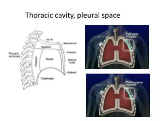
Pneumothorax is defined as the presence of air within the pleural cavity.
Pneumothorax. Spontaneous Primary spontaneous Secondary spontaneous Traumatic Non-iatrogenic Iatrogenic Accidental Artificial
Causes of Pneumothorax . Spontaneous pneumothorax Primary apical blebs stature Secondary Spontaneous Pneumothorax chronic bronchitis and emphysema asthma suppurative pneumonia tuberculosis of the lungs cystic fibrosis
Traumatic pneumothorax Non-iatrogenic open and closed chest injury barotrauma Iatrogenic (accidental) paracentesisthoracis pleural biopsy transbronchial biopsy percutaneous lung biopsy/aspiration central venous cannulation barotrauma (mechanical ventilation) Iatrogenic (deliberate) artificial pneumothorax
Radiographic appearances Characteristic finding is a sharply defined lung edge that is convex outwards and separated from the chest wall by a lucent zone entirely devoid of lung marking. If the pneumothorax is shallow: Chest radiograph is taken in full expiration. Other method is that the chest radiograph is taken in the lateral decubitus position with the side on which the pneumothorax is suspected the upper most. In tension pneumothoraxthe lung may be compressed into a shapeless shadow or even displaced across the midline along with the mediastinal structures.
A 2 cm rim of air actually represents approximately 50% of the hemithoracic volume.
Primary Pneumothorx Small(stable) Observe 3 _ 6 hours. Repeat CXR (no increase in size ) disharge. Advice to come after 12 to 48 with fresh X- ray chest. There is 1.25 % re-expansion of hemi thorax volume per day
LARGE PNEUMOTHORAX(stable) Hospitalize . Aspirate with 14 F cannula. Tube Thorocostomy
LARGE PNEUMOTHORAX ( unstable ) Hospitalize. Tube Thorocostomy. Suction (5 – 15 cm of water ). Persistent leak ( 4 – 6 week ) Ref. to Thoracic Surgeon.